
Method for Image Outlier Removal for Transmission Electron Microscope Cameras

a transmission electron microscope and image processing technology, applied in image enhancement, immunological disorders, antibody medical ingredients, etc., can solve the problems of not fully effective, method is not fully effective, and only partially effective at performing this latter function

Active Publication Date: 2015-11-19
GATAN INC
View PDF1 Cites 3 Cited by
  • Summary
  • Abstract
  • Description
  • Claims
  • Application Information

AI Technical Summary

Benefits of technology

The patent text describes a method for removing outliers from images captured by electron microscopes. These outliers are caused by X-rays and other sources of radiation that can damage the sensor and affect image quality. The method uses image processing techniques to identify and remove these outliers, but there are problems with existing methods, such as the inability to effectively shield X-rays and the use of a single threshold for identifying outliers. The patent proposes a new method that addresses these issues and can effectively remove outliers from high-contrast or rapidly varying specimen images.

Problems solved by technology

The optic is only partially effective at performing this latter function.
These spots do not contribute to the quality of the image but, rather, detract from it because 1) the random emission angle at the scintillator and subsequent scattering destroy any spatial correlation of the x-rays with the image-signal-bearing electrons which created them at the scintillator and 2) because the directly detected x-ray adds significantly more signal to the image detected at the silicon sensor than the net signal conferred by the incoming electron after scintillation and image transfer by the optic.
This method is not fully effective if either the energy of the electron beam is too high or if there is not sufficient room to place enough material between scintillator and sensor.
Furthermore, in the case of lens optics, the optical design will dictate the amount of material which might not be sufficient to provide adequate shielding of x-rays.
Shielding by the camera housing and by the optics serves to reduce these forms of radiation as well but, as in the case of bremsstrahlung, this is not 100% effective.
In addition, direct detection sensors without a scintillator and transfer optics will have issues with detection of stray scatter and other radiation sources which are not described by the statistics of the specimen image and which are disturbing to the image quality.
There are a number of problems with this approach.
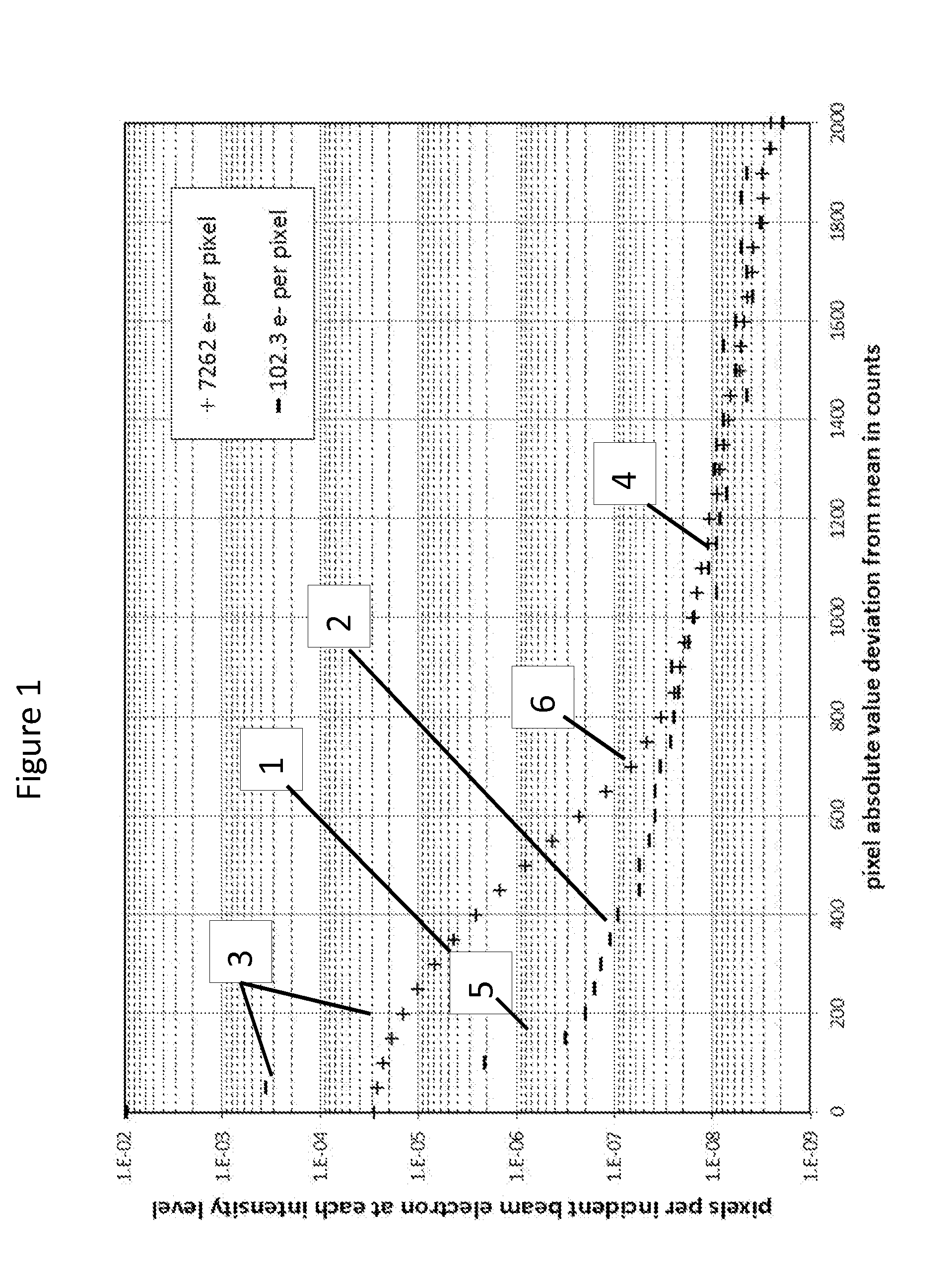
A second problem is that as dose level increases, the histogram of ordinary Gaussian noise in the indirect image (the intended, “good” image from scintillator light generation, optical transfer and sensor detection) grows to obscure more and more of the histogram of the directly detected radiation events.
For high-contrast images with significant spatial variation, errors can be introduced here as well, in that average-setting and interpolation will both be inaccurate if the image is too rapidly varying as a function of position.

Method used


Image

  • Method for Image Outlier Removal for Transmission Electron Microscope Cameras
  • Method for Image Outlier Removal for Transmission Electron Microscope Cameras
  • Method for Image Outlier Removal for Transmission Electron Microscope Cameras

Examples


Embodiment Construction

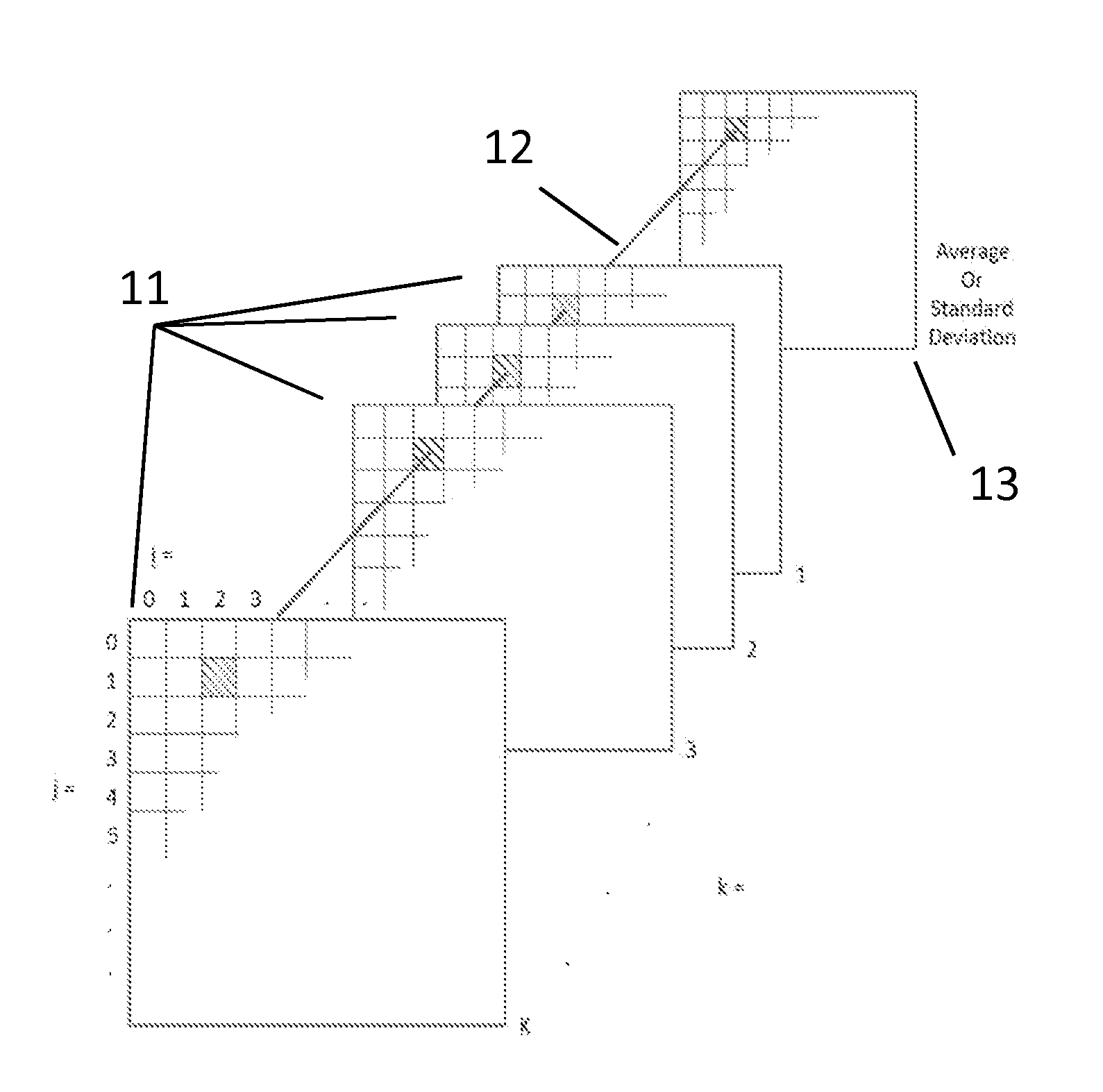
[0020]Fractionating the dose of an exposure, i.e. dividing it into a series of sub-exposures which still add up to the same total dose, creates a number of opportunities to improve outlier removal. First, it allows the evaluation of mean and standard deviation for the determination of an outlier threshold on a pixel-by-pixel basis by providing an exposure series for each pixel and thereby avoids reliance of the method on the assumption of ergodicity. In turn, by virtue of making the statistical evaluation pixel-wise, it largely removes the sensitivity to specimen image characteristics. Second, by reducing the dose at which thresholding is done, less of the direct radiation detection histogram is subsumed into the indirect image signal allowing choice of a lower threshold and a more complete removal of unintended radiation signal. Third, since each sub-exposure can be corrected individually, each pixel of the final summed image will have reduced artifacts from replacement of outliers...


PUM

PropertyMeasurementUnit
Threshold limitaaaaaaaaaa

Abstract

Methods are disclosed for removal of outlier pixels from a transmission electron microscopy camera image. One exemplary method includes establishing a desired exposure of n electrons per pixel; exposing the camera to a series of sub-frame exposures to produce a series of sub-frame images; calculating an average image signal of all sub-frame exposures in said series; establishing a threshold selected to achieve a desired number of false positives; evaluating each of said sub-frame exposures for pixels further away from said average than said threshold; and replacing pixels in each of said sub-frame images that exceed said threshold with said average to form corrected sub-frame images.

Description

CROSS REFERENCE TO RELATED APPLICATION[0001]This utility application claims the benefit under 35 U.S.C. §119(e) of provisional application Ser. No. 60 / 032,535 filed on Aug. 2, 2014, and entitled “Method for Image outlier Removal for Electron Microscope Cameras.” The entire disclosure of the provisional application is incorporated herein by reference.FIELD OF THE INVENTION[0002]The invention relates generally to the field of electron microscopy image processing.BACKGROUND OF THE INVENTION[0003]Until recently cameras for electron microscopes have been manufactured exclusively using an indirect imaging method employing either a scintillator to convert an electron image into a light image, some form of optics to capture the light image and transfer it to a plane offset from the scintillator, and a silicon sensor to capture the light image. The optics have been composed either of a fused fiber optic plate or of a lens. Aside from the primary function of light image transfer, the optics a...

Claims


Application Information

Patent Timeline
IPC IPC(8): H01J37/26C12N15/09A61K39/00A61P37/04A61P43/00C12N5/00C12N5/10
CPCH01J37/263G06T2207/10061H04N25/683G06T5/77A61K39/00H01J37/222H01J37/26H01J2237/223H01J2237/226H01J2237/2448H01J2237/24495H01J2237/2617A61P37/04A61P43/00A61K48/00C12N15/09
Inventor MOONEY, PAUL
Owner GATAN INC
Learn More