PD-L1 Antibodies and Uses Thereof

a technology of pdl1 and antibodies, applied in the field of pdl1 antibodies, can solve the problems of unreliable diagnostic reagents, conflicting results, and challenging use of pdl1 protein expression as an accurate predictor of cancer

Active Publication Date: 2015-12-03
VENTANA MEDICAL SYST INC
View PDF2 Cites 57 Cited by
  • Summary
  • Abstract
  • Description
  • Claims
  • Application Information

AI Technical Summary

Benefits of technology

[0038]In one aspect, provided herein is a method of detecting PD-L1 in a tumor sample comprising (a) contacting the sample with an antibody or an antigen binding fragment of the antibody, wherein the antibody comprises a heavy chain (HC) immunoglobulin variable domain sequence and a light chain (LC) immunoglobulin variable domain sequence, wherein the antibody binds to an epitope of human PD-L1 comprising the amino acid sequence CGIQDTNSKKQSDTHLEET (SEQ ID NO: 1) and/or has a half maximal effective concentration (EC50) of at least 1.5×10⁻¹¹ M, wherein the HC comprises (i) a HC CDR1 comprising the amino acid sequence NHAIS (SEQ ID NO: 14); (ii) a HC CDR2 comprising the amino acid sequence TINSDTHTYYATWPKG (SEQ ID NO: 15); and (iii) a HC CDR3 comprising the amino acid sequence RIFSSSNI (SEQ ID NO: 16); and the LC co

Problems solved by technology

However, the use of PD-L1 protein expression as an accurate predictor for cancer and/or the efficacy of anti-PD-1 and anti-PD-L1 directed therapies remains challenging.
Many commercially available antibodies directed to PD-L1 cross-react with other protein

Method used


Image

  • PD-L1 Antibodies and Uses Thereof
  • PD-L1 Antibodies and Uses Thereof
  • PD-L1 Antibodies and Uses Thereof

Examples


example 1

Rabbit Monoclonal Antibody Generation

[0201]FIG. 1 illustrates the overall procedure used to create PD-L1 monoclonal antibodies using a rabbit host. The anti-PD-L1 rabbit monoclonal primary antibodies were directed against the sequence CGIQDTNSKKQSDTHLEET (SEQ ID NO: 1), which represents amino acid residues 272-290 of human PD-L1. Thus, the resulting antibodies would target the C-terminal cytoplasmic region of human PD-L1 much like the E1L3N® anti-PD-L1 antibody (Cell Signaling Technology, MA).

[0202]The 19-amino acid peptide was synthesized and covalently conjugated to a keyhole limpet haemocyanin (KLH) carrier protein. New Zealand white rabbits were immunized with KLH-conjugated peptide emulsified with complete Freund's adjuvant followed by a series of booster doses of immunogen emulsified with incomplete Freund's adjuvant. The rabbit that generated a IHC positive polyclonal antibody was selected for further monoclonal development. For IHC testing, standard OptiView DAB kit protocol...

example 2

Target Specificity of Anti-PD-L1 Antibodies

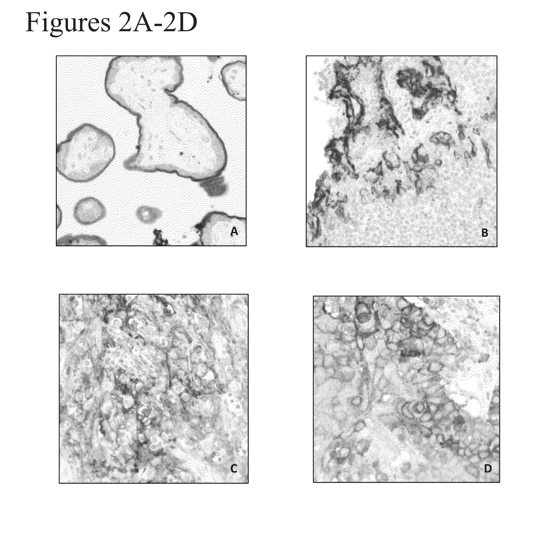
[0212]Rabbit anti-human PD-L1 monoclonal antibodies SP263, J45H2L4 and J27H6L4 were applied onto formalin-fixed paraffin embedded (FFPE) tissue samples to assess the staining patterns of these antibodies. Tissue samples include placenta (positive control), stomach (negative control) and colon. Immunohistochemistry was performed on BenchMark Ultra (Ventana Medical System) using StdCC1 cell conditioning and standard Opt iView DAB detection protocol. Each primary antibody was incubated at 0.9 μg / ml for 16 min.

[0213]As shown in FIG. 4G, the J27H6L4 anti-PD-L1 antibody exhibited weak staining in placental trophoblasts and no staining in colon and stomach tissue (FIGS. 4H-4I). In contrast, the J45H2L4 anti-PD-L1 antibody generated the strongest signal in placental trophoblasts. See FIG. 4D. However, J45H2L4 also exhibited significant background staining, as demonstrated by the non-specific nuclear staining in colon and stomach tissue (FIGS. 4E an...

example 3

Characterization of the SP263 Anti-Human PD-L1 Antibody

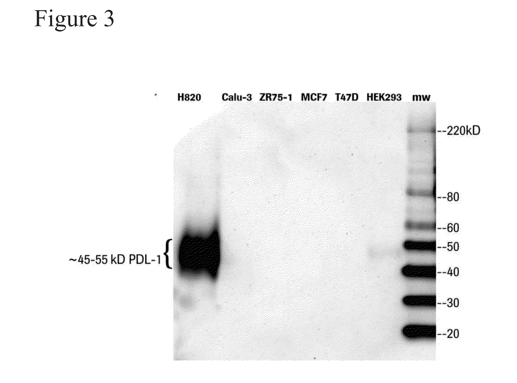
[0216]Western blot analysis was used to assess the binding specificity of the SP263 anti-PD-L1 antibody in biological samples. Cell lysates from a NIH H820 lung adenocarcinoma cell line (positive control), a HEK293 cell line, a Calu-3 lung adenocarcinoma cell line (negative control), a ZR75-1 human breast carcinoma cell line (negative control), a MCF7 human breast carcinoma cell line (negative control), and a T47D human breast carcinoma cell line (negative control) were fractionated by SDS-PAGE and was subjected to western blotting with the SP263 anti-PD-L1 antibody using standard techniques (see OptiView DAB detection protocol).

[0217]As shown in FIG. 3, SP263 bound a ˜45-55 kDa protein which corresponds to human PD-L1 protein. The 45-55 kDa PD-L1 protein was detected in NIH H820 lung adenocarcinoma cells (which are known to exhibit elevated levels of PD-L1), and was absent in all 4 negative controls. Further, in addition to spe...


PUM

PropertyMeasurementUnit
Solutionaaaaaaaaaa

Abstract

Provided herein are novel PD-L1 antibodies and methods for using the same for diagnosing a medical condition associated with elevated PD-L1 levels (e.g., cancer) in subjects in need thereof and antigen binding fragments thereof. The PD-L1 antibodies and antigen binding fragments are also useful in evaluating the efficacy of a particular therapeutic regime in a subject diagnosed as having a PD-L1-related medical condition.

Description

CROSS-REFERENCE TO RELATED APPLICATIONS[0001]This disclosure claims the benefit of U.S. 62 / 004,572, filed May 29, 2014, and U.S. 62 / 069,420, filed Oct. 28, 2014, the contents of which are hereby incorporated by reference in their entirety.BACKGROUND OF THE INVENTION[0002]1. Field of the Invention[0003]This disclosure relates to novel PD-L1 antibodies and methods for using the same for detecting PD-L1 polypeptides in a biological sample. PD-L1 antibodies also are useful to evaluate the efficacy of a particular therapeutic agent in a subject diagnosed as having a PD-L1-related medical condition.[0004]2. Description of Related Art[0005]The following description is provided to assist the understanding of the reader. None of the information provided or references cited is admitted to be prior art.[0006]Programmed death 1 (PD-1) is a member of the CD28 family of receptors, which includes CD28, CTLA-4, ICOS, PD-1, and BTLA. The initial members of the family, CD28 and ICOS, were discovered ...

Claims


Application Information

Patent Timeline
IPC IPC(8): G01N33/574C07K16/28
CPCG01N33/57492C07K16/2896G01N2333/70596C07K2317/51C07K2317/515C07K2317/92C07K16/2827C07K2317/34G01N33/574
Inventor COUTO, FERNANDO JOSE REBELO DOLIAO, ZHIMINGZHU, YIFEI
Owner VENTANA MEDICAL SYST INC
Why Eureka
Try Eureka