Determining a Three-dimensional Model Dataset of a Blood Vessel System with at least One Vessel Segment

a blood vessel system and three-dimensional model technology, applied in image data processing, diagnostics, applications, etc., can solve the problems of manual segmentation of projection images, difficult recording of blood vessel images, and relatively complex specification of the approximate vessel center line of a relevant vessel. achieve the effect of simple and reliable registration of projection images

Active Publication Date: 2016-01-14
SIEMENS HEALTHCARE GMBH
View PDF6 Cites 5 Cited by
  • Summary
  • Abstract
  • Description
  • Claims
  • Application Information

AI Technical Summary

Benefits of technology

[0030]In particular, branches of the blood vessel system may be used as landmarks. The landmarks of different projection images may be assigned to one another as a function of the classification information of the image areas lying in the area of the landmarks. In particular, through the assignment of the different classification information to the image areas immediately adjoining one another in the different projection images, t

Problems solved by technology

Recording images of the blood vessel system is also made more difficult by the fact that vessels in the heart region move a great deal.
Tomographic image reconstruction is typically not possible under these conditions.
The manual segmentation of projection images, as well as th

Method used


Image

  • Determining a Three-dimensional Model Dataset of a Blood Vessel System with at least One Vessel Segment
  • Determining a Three-dimensional Model Dataset of a Blood Vessel System with at least One Vessel Segment
  • Determining a Three-dimensional Model Dataset of a Blood Vessel System with at least One Vessel Segment

Examples


Embodiment Construction

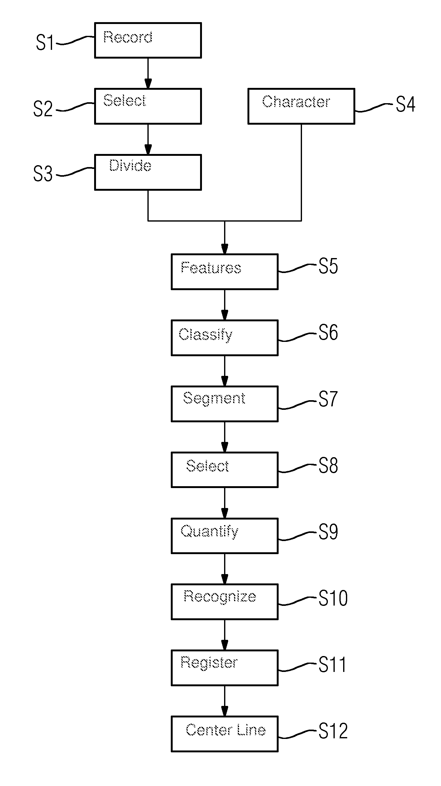
[0049]FIG. 1 shows an exemplary embodiment of the method for determining a three-dimensional model dataset of a blood vessel system of a patient including at least one vessel segment. In act S1, as a preparatory act, a number of projection images of a blood vessel system of a patient are recorded from different recording angles with an imaging detection device, namely an x-ray device with a C-arm. In particular, a contrast medium is used during the recording of the blood vessel system. The recording angles of the projection images are selected such that an angular range of at least 30° is covered by the projection images. The recording is triggered by a synchronization of the recordings with an EKG signal such that all recordings occur in the same heart phase, especially during the end systole or the end diastole. A heart phase is selected in which a small movement in the blood vessel system, even in the area adjacent to the heart, is to be expected.

[0050]In act S2 the projection im...


PUM


Abstract

A three-dimensional model dataset of a blood vessel system of a patient including at least one the vessel system is determined from a number of projection images, which have been recorded from different recording angles, of the blood vessel system The projection images are divided up into image areas each containing at least one pixel. A feature vector is determined for each of the image areas. Classification information, which describes how the respective image area belongs or does not belong to a vessel segment of the blood vessel system defined in accordance with anatomical specification data, is defined for each of the image areas by applying a classification function to the feature vector assigned to the image area. The classification function has been trained by training data records annotated with classification information obtained from at least one person other than the patient. The blood vessel system in the projection images is segmented by grouping image areas with the same classification information. The three-dimensional model dataset is calculated as a function of the segmented projection images and the classification information.

Description

RELATED CASE[0001]This application claims the benefit of DE 102014213408.0, filed on Jul. 10, 2014, which is hereby incorporated by reference in its entirety.FIELD[0002]The embodiments relate to a method for determining a three-dimensional model dataset of a blood vessel system including at least one vessel segment of a patient.BACKGROUND[0003]It is advantageous, particularly for planning and monitoring of catheter interventions, to use a three-dimensional model of a blood vessel system of a patient. Such a three-dimensional presentation is mostly intended to be reconstructed from a number of two-dimensional image recordings, especially x-ray recordings. In principle, it would be possible in such cases to calculate a 3D presentation from a number of two-dimensional projection images of vessels, especially with administration of contrast media. In everyday clinical practice, however, additional requirements are to be placed on the imaging of the blood vessel system. Thus, images are ...

Claims


Application Information

Patent Timeline
IPC IPC(8): G06T17/10A61B6/00A61B6/03G06T11/60
CPCG06T17/10G06T11/60A61B6/03G06T2200/04A61B6/5217A61B6/504A61B6/5294A61B6/032A61B6/4441A61B6/5205A61B6/5288G06T17/00G06T2210/41
Inventor LAURITSCH, GUNTERREDEL, THOMASROHKOHL, CHRISTOPHERSCHEUERING, MICHAEL
Owner SIEMENS HEALTHCARE GMBH
Why Eureka
Try Eureka