In vitro biliary excretion assay
- Summary
- Abstract
- Description
- Claims
- Application Information
AI Technical Summary
Benefits of technology
Problems solved by technology
Method used
Image
Examples
example 1
Hepatocyte-Stromal Cell Cocultures
[0131]Cryopreserved human hepatocytes were removed from liquid nitrogen and thawed. After thawing, cells were resuspended in medium and cell number and cell viability was determined using trypan blue exclusion. Stromal cells were passed in a CO2 incubator until used for experimental plating. On plating day cells were detached from the plate, washed, and resuspended in medium. Cell number and viability were determined using trypan blue exclusion.
[0132]Hepatocytes and stromal cells were seeded into collagen-coated 96-well plates at a density of 30,000 hepatocytes per well. The stromal cells were growth arrested prior to seeding. Cultures were maintained for 7 days before the start of any biliary excretion assays. Before the experiments were started, the cells were stained with CDFDA on day 7 to ensure canalicular formation.
example 2
Excretion of Test Compound Into Bile Canaliculi in Hepatocyte-Stromal Cell Cocultures
[0133]In this experiment the presence of bile canaliculi in hepatocyte-stromal cell cocoultures prepared according to Example 1 was assessed. Cocultures were incubated with 5 uM 5-(and-6)-Carboxy-2′,7′-dichloro-fluoreceine diacetate (CDFDA) for 20 min. CDFDA that is taken up by hepatocytes is hydrolyzed to fluorescent 5-(and-6)-Carboxy-2′,7′-dichloro-fluoreceine (CDA) by the hepatocyte and then secreted in this fluorescent form into the canaliculus via the Mrp2 transporter. The cocultures comprised several canaliculi that are readily apparent under the microscope (data not shown). Cyclosporin A (CSA) is a known inhibitor of the Mrp2 transporter. In a subsequent experiment a coculture was incubed with CDFDA in the presence of 50 uM CSA for 20 min. No fluorescent canaliculi were visible following incubation with CDFDA in the presence of CSA (data now shown). This result demonstrates that bile canalicu...
example 3
Disruption of Canaliculi
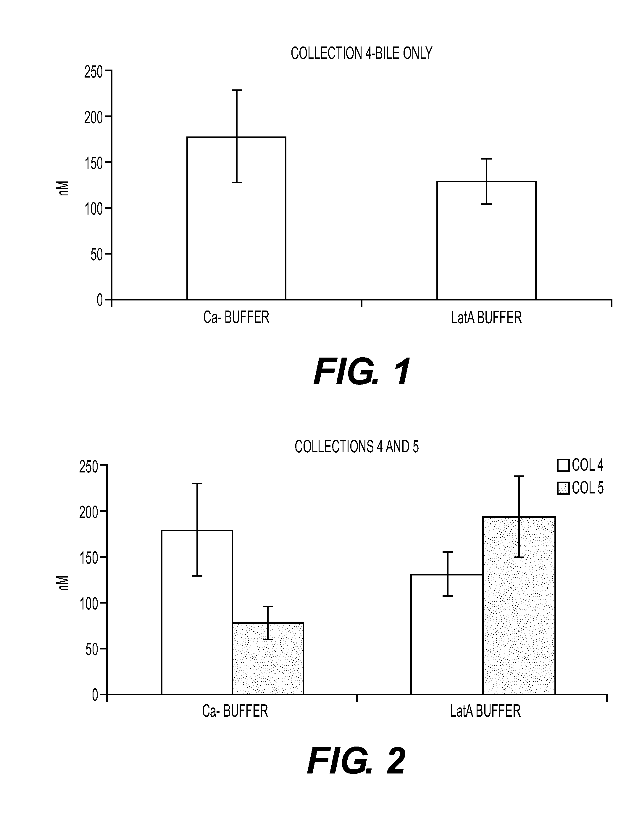
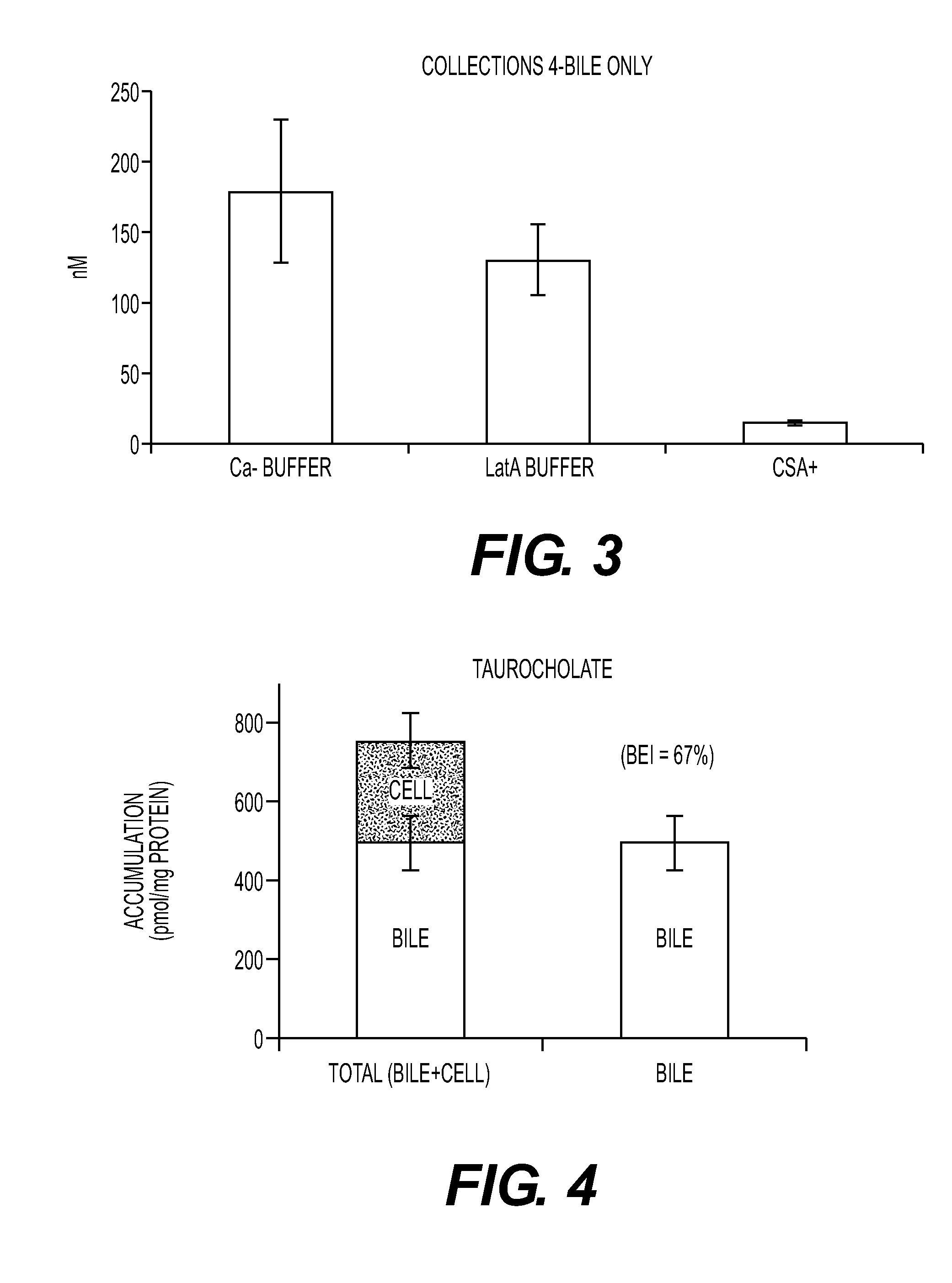
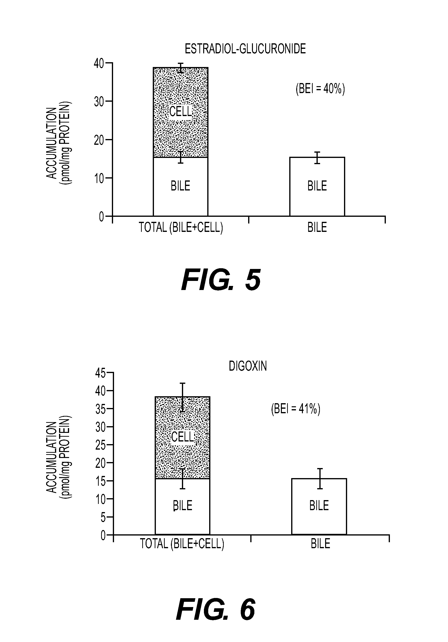
[0134]Assessing excretion of a chemical entity into the bile in the hepatocyte-stromal cell coculture requires a method of distinguishing between the chemical entity present in canaliculi of the coculture and chemical entity that may be present in the cytoplasm of cells in the coculture. Two methods of canalicular disruption were tested. In the first method, the cultures were exposed to a calcium free (Ca−) free buffer. The lack of calcium inhibits the ability of the cell to form tight junctions which allows bile present in the canaliculi to escape. Cells were first washed two times in HBSS buffer with Ca+. Cells were then incubated with 5 uM CDFDA in HBSS buffer without Ca− for 20 min. Cells were then washed two times in HBSS buffer without Ca−. Because accumulated CDF escaped the canaliculi after exposure to Ca− buffer no CDF staining was apparent (data not shown).
[0135]In the second method, the cultures were exposed to buffer containing latranculin A (LatA...
PUM
 Abstract
Description
Claims
Application Information
 - R&D
- Intellectual Property
- Life Sciences
- Materials
- Tech Scout
- Unparalleled Data Quality
- Higher Quality Content
- 60% Fewer Hallucinations
Browse by: Latest US Patents, China's latest patents, Technical Efficacy Thesaurus, Application Domain, Technology Topic, Popular Technical Reports.
© 2025 PatSnap. All rights reserved.Legal|Privacy policy|Modern Slavery Act Transparency Statement|Sitemap|About US| Contact US: help@patsnap.com


