Handheld and portable scanners for millimeter wave mammography and instant mammography imaging

a millimeter wave and instant mammography technology, applied in the field of sensor and imaging systems, can solve the problems of large, not portable, bulky system, generally requires a moderately expensive system,

Active Publication Date: 2016-03-10
MOHAMADI FARROKH
View PDF2 Cites 8 Cited by
  • Summary
  • Abstract
  • Description
  • Claims
  • Application Information

AI Technical Summary

Problems solved by technology

X-ray mammography, which requires patient contact with the scanner and ionizing radiation impact on the patient, may produce 7% false positives, as high as 40% false negatives, and generally requires a bulky (large, not portable) system that is typically expensive.
Infrared imaging, which may not require patient contact with the scanner, relies on thermology based on blood vessels (neo-angiogenesis); disadvantages include a resolution greater than 12 millimeters (mm), shallow depths of detection, may produce 19% false positives, and generally requires a moderately expensive system.
Elastic-scattering spectroscopy requires contact with the patient, employing an optical probe biopsy; an injection is required, leading to issues with dispersion for imaging.
Scintimammography uses injection of a tracer (a radioactive chemical) that gets collected inside a tumor; disadvantages are seen in the use of radioactive material in a lengthy process resulting in resolution of only 9 mm, and generally requires an expensive system.
Ultrasound probes can identify various types of lumps, such as liquid-filled cysts and solid masses, but requires contact with the patient, a lengthy process resulting in resolution of 3 mm, a high false positive rate, and typically requires an expensive system.
Confocal microwave imaging employs an array of antennas placed around the breast being scanned requiring contact with the patient, resulting in resolution of 3 mm, and generally requires a bulky (large, not portable) system.
Positron emission tomography (PET) employs radioactive glucose that is injected into the patient's body, which is then scanned, and can result in higher doses of X-ray radiation (e.g., 20-30 times that of X-ray mammography); and generally requires a bulky and expensive system.
Magnetic resonance imaging (MRI) does not require patient contact and may be more suitable for dense breast tissues; but is prone to false positives, and generally requires a bulky and expensive system.

Method used


Image

  • Handheld and portable scanners for millimeter wave mammography and instant mammography imaging
  • Handheld and portable scanners for millimeter wave mammography and instant mammography imaging
  • Handheld and portable scanners for millimeter wave mammography and instant mammography imaging

Examples


Embodiment Construction

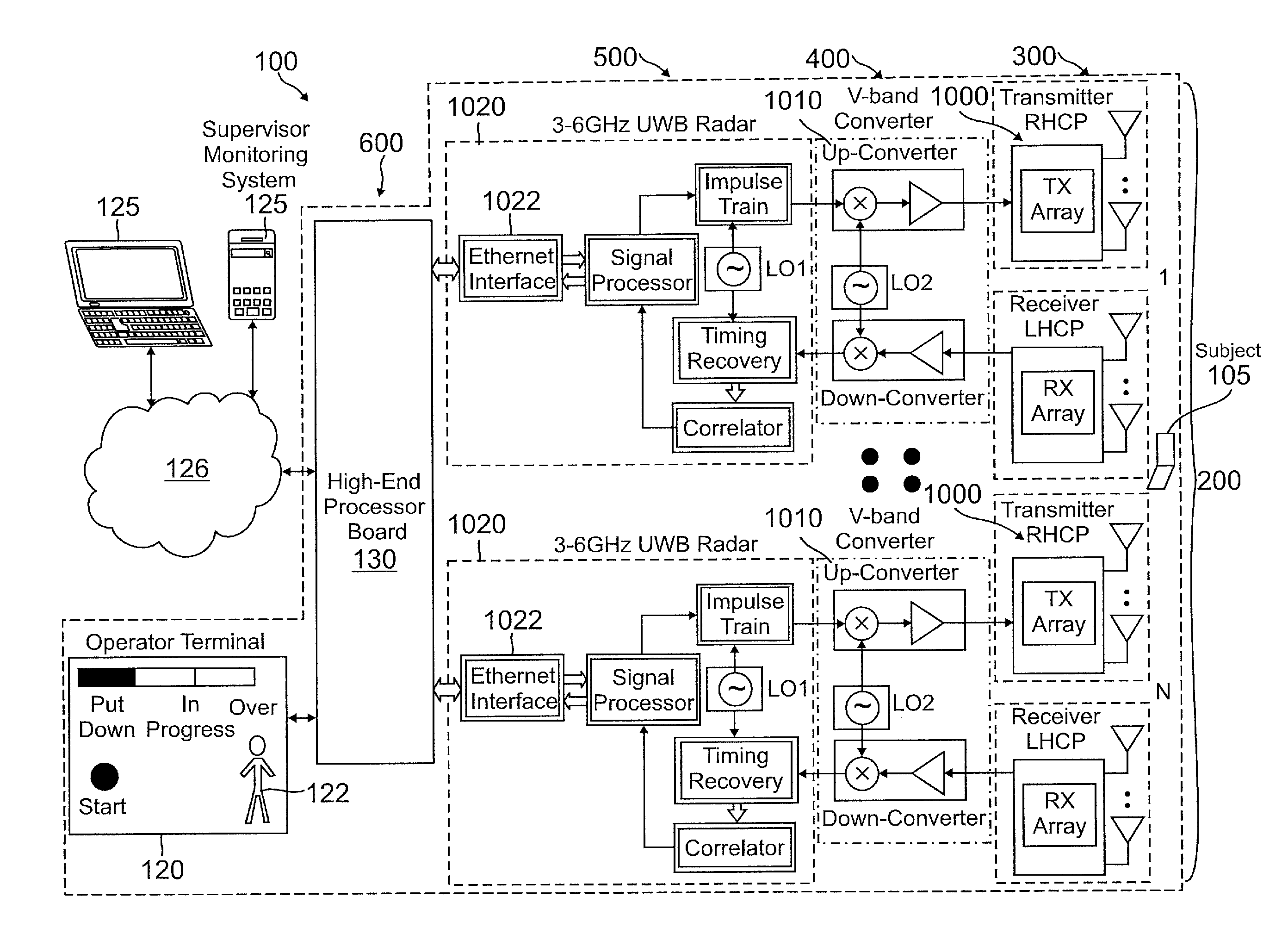
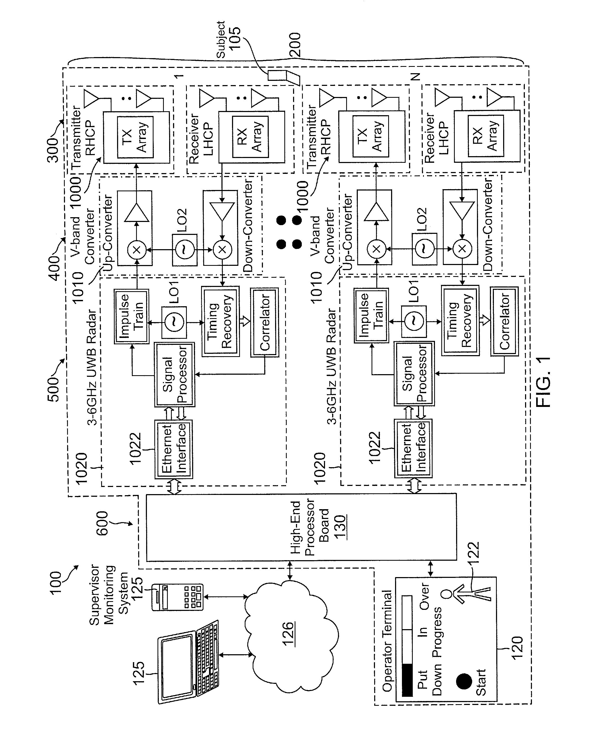
[0023]Embodiments of the disclosure address the need for a handheld or portable scanning device and real-time imaging that can be used, for example, in a primary care physician's office. Methods and systems are disclosed for ultra wide band (UWB) sensor imaging for medical diagnosis that address the needs for early detection of cancer that does not require patient contact or injections, does not expose the patient to unsafe radiation, does not require painful compression of breast tissue such as experienced with conventional X-ray mammography, does not require a high level of skill on the part of the operator to obtain accurate scans while ensuring patient safety, and provides a low cost, portable or handheld, real-time imager that can be used, for example, in a primary care physician's office. In one embodiment, mobile, handheld, and versatile performance can be obtained in any office or even at home on account of the small volume of the sensor and imaging units which can be on the...


PUM


Abstract

Methods and systems provide a non-ionizing alternative to conventional mammography X-ray techniques, which expose patients to ionizing radiation, for breast cancer tumor detection, using a miniaturized (wafer scale) array of ultra wide band (UWB) radio frequency (RF) sensors operating at 60 GHz (non-ionizing—no X-ray type accumulative radiations) that have capability to use both linear and polarized sensors, tomography, and suppression of scattering for improved imaging. Coding techniques provide significant processing gain that is essential for the large attenuation of transmitted signals in breast tissue operating at these high frequencies. The increased bandwidth of UWB RF detection provides better depth resolution of breast and body tissue. Using polarization improves detection of abnormal tissues. An extremely miniaturized (wafer scale) cluster of transmitter and receiver antenna elements improves detection at deeper parts of the breast and can detect cancerous cells in dense breasts often not picked up by mammography.

Description

CROSS REFERENCE TO RELATED APPLICATIONS[0001]This application claims the benefit of priority from U.S. Provisional Patent Application No. 62 / 046,826, filed Sep. 5, 2014, which is incorporated by reference.BACKGROUND[0002]1. Field of the Invention[0003]The present invention relates generally to sensor and imaging systems and, more particularly, to ultra wideband sensor systems integrated with wafer scale antenna arrays for tissue imaging and cancer diagnosis in the field of medicine.[0004]2. Related Art[0005]The incidence of cancer and, more particularly, that of breast cancer, is generally recognized as a serious medical, economical, and human problem. That the financial costs of cancer can be high for both the person with cancer and for society as a whole is illustrated by the following statistics from the National Institutes of Health (NIH), which estimated the 2009 overall annual costs of cancer were as follows: 1) in 2009: new cases of breast cancer: 173,400, deaths from breast ...

Claims


Application Information

Patent Timeline
Patent Type & Authority Applications(United States)
IPC IPC(8): A61B5/05A61B5/00
CPCA61B5/0507A61B2576/02A61B5/7246A61B5/4312A61B2560/0431G16H30/40
Inventor MOHAMADI, FARROKH
Owner MOHAMADI FARROKH
Why Eureka
Try Eureka