Method and system for automated detection and measurement of a target structure

a target structure and automatic detection technology, applied in the field of diagnostic imaging, can solve the problems of complex acquisition of optimal image frame that includes clinically prescribed scan plane for satisfying prescribed clinical guidelines, confusion of bpd and hc measurements, and difficulty in achieving optimal image frame acquisition

Inactive Publication Date: 2016-03-24
GENERAL ELECTRIC CO
View PDF5 Cites 32 Cited by
  • Summary
  • Abstract
  • Description
  • Claims
  • Application Information

AI Technical Summary

Problems solved by technology

Acquisition of an optimal image frame that includes the clinically prescribed scan plane for satisfying prescribed clinical guidelines, however, may be complicated.
For example, acquisition of the optimal image frame may be impaired due to imaging artifacts caused by shadowing effect of bones, near field haze resulting from subcutaneous fat layers, unpredictable patient movement, and / or ubiquitous speckle noise.
Additionally, an unfavorable fetal position, fetus orientation, and / or change in shape of the fetal head due to changes in the transducer pressure may also confound the BPD and HC measurements.
Moreover, operator and / or system variability may also limit reproducibility of the BPD and HC measurements.
Additionally, sub-optimal ultrasound image settings such as gain compensation and dynamic range may impede an ability to visualize internal structures of the human body.
Furthermore, even small changes in positioning the ultrasound transducer may lead to significant changes in the visualized image frame, thus leading to incorrect measurements.
While experienced radiologists may be able to obtain accurate measurements with less effort and time, acquiring clinically acceptable biometric measurements typically requires much greater effort and time from inexperienced users and / or entails use of expensive 3D ultrasound probes.
Accordingly, accuracy of conventional ultrasound imaging methods may depend significantly upon availability of state-of-the-art ultrasound probes and / or skill and experience of the radiologist, thereby limiting availability of quality imaging services, for example, to large hospitals and urban areas.
Scarcity of skilled and / or experienced radiologists in remote or rural regions, thus, may cause these regions to be poorly or under-served.
However, upon failing to find a better image frame, the radiologist may have to manually scroll back to an originally acceptable image frame, thus prolonging imaging time and hindering reproducibility.
Ultrasound imaging using conventional methods, cost-effective ultrasound scanners, and / or by a novice radiologist, therefore, may not allow for measurements suited for real-time diagnosis and treatment.

Method used


Image

  • Method and system for automated detection and measurement of a target structure
  • Method and system for automated detection and measurement of a target structure
  • Method and system for automated detection and measurement of a target structure

Examples


Embodiment Construction

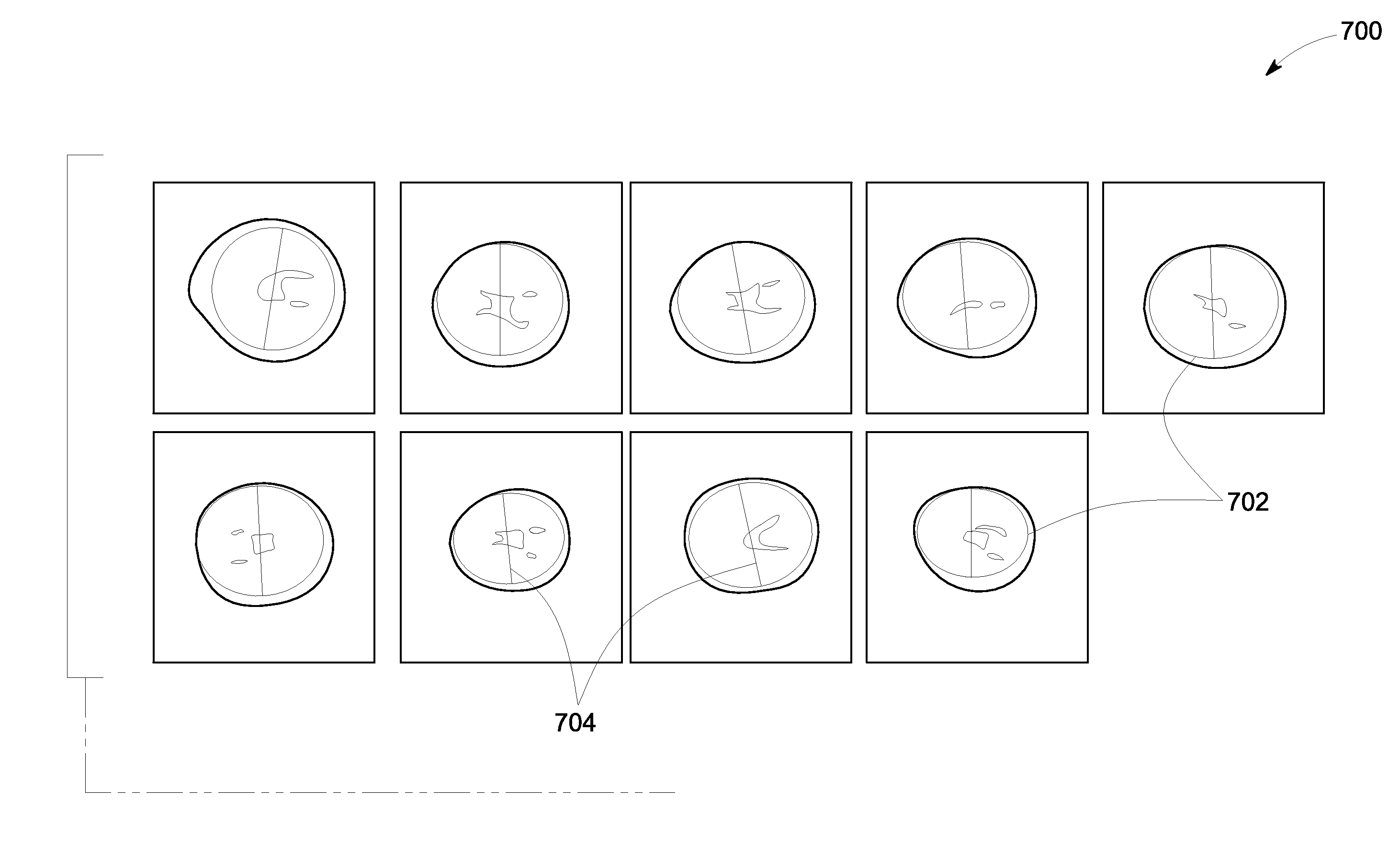
[0018]The following description presents systems and methods for automatically detecting and measuring a target structure in an ultrasound image. Particularly, certain embodiments presented herein describe the systems and methods configured to accurately detect one or more target structures in a plurality of image frames and identify an optimal image frame that includes the one or more target structures in a desired scan plane. As used herein, the term “desired scan plane” may be used to refer to a cross-sectional slice of an anatomical region that satisfies clinical, user-defined, and / or application-specific guidelines to provide accurate and reproducible measurement of a target structure. Furthermore, the term “optimal image frame” is used to refer to an image frame that includes the target structure in the desired scan plane that satisfies the prescribed guidelines for providing one or more desired measurements of the target structure.

[0019]Particularly, the target structure, for...


PUM


Abstract

A system and method for imaging a subject are disclosed. A plurality of edge points corresponding to a set of candidate structures are determined in each image frame in a plurality of 3D image frames corresponding to a volume in the subject. A target structure is detected from the set of candidate structures by applying constrained shape fitting to the edge points in each image frame. A subgroup of image frames including the target structure is identified from the 3D frames. A subset of edge points corresponding to the target structure is determined in each of the subgroup of image frames. A plurality of 2D scan planes corresponding to the subset of edge points is determined, and ranked using a determined ranking function to identify a desired scan plane. A diagnostic parameter corresponding to the target structure is measured using a selected image frame that includes the desired scan plane.

Description

BACKGROUND[0001]Embodiments of the present specification relate generally to diagnostic imaging, and more particularly to a method and system for automatically detecting and measuring a target structure in an ultrasound image.[0002]Medical diagnostic ultrasound is an imaging modality that employs ultrasound waves to probe the acoustic properties of biological tissues and produces corresponding images. Particularly, ultrasound systems are used to provide an accurate visualization of muscles, tendons, and other internal organs to assess their size, structure, and any pathological conditions using near real-time images. For example, ultrasound images have been extensively used in prenatal imaging for assessing gestational age (GA) and weight of a fetus. In particular, two-dimensional (2D) and / or three-dimensional (3D) ultrasound images are employed for measuring desired features of the fetal anatomy such as the head, abdomen, and / or femur. Measurement of the desired features, in turn, ...

Claims


Application Information

Patent Timeline
Patent Type & Authority Applications(United States)
IPC IPC(8): A61B8/08A61B5/00A61B8/00G06T7/00
CPCA61B8/5223G06T7/0014G06T7/0085G06T7/0089A61B8/483A61B2576/00A61B8/0866A61B8/4245G06T2207/10136G06T2207/20124G06T2207/30044A61B5/0073A61B5/06A61B8/0841A61B8/085A61B8/0875A61B8/4254A61B8/466A61B8/469A61B8/481A61B8/5207G06T2207/20081A61B5/065A61B5/067A61B5/1076A61B2503/02G06T7/73G06T7/12G06T7/62G16H50/30G06V10/7515G06V2201/03
Inventor CHEN, JIXUKRISHNAN, KAJOLI, BANERJEE
Owner GENERAL ELECTRIC CO
Why Eureka
Try Eureka