

System and method for detecting abnormalities in cervical cells
a technology of cytologic examination, which is applied in the field of system and method for detecting abnormalities in human cervical and vaginal cells, can solve the problems of poor reproducibility, limited positive predictive value of hpv testing, and relatively insensitive single cytologic examination
- Summary
- Abstract
- Description
- Claims
- Application Information
AI Technical Summary
Benefits of technology
Problems solved by technology
Method used
Image

Examples
specific embodiments
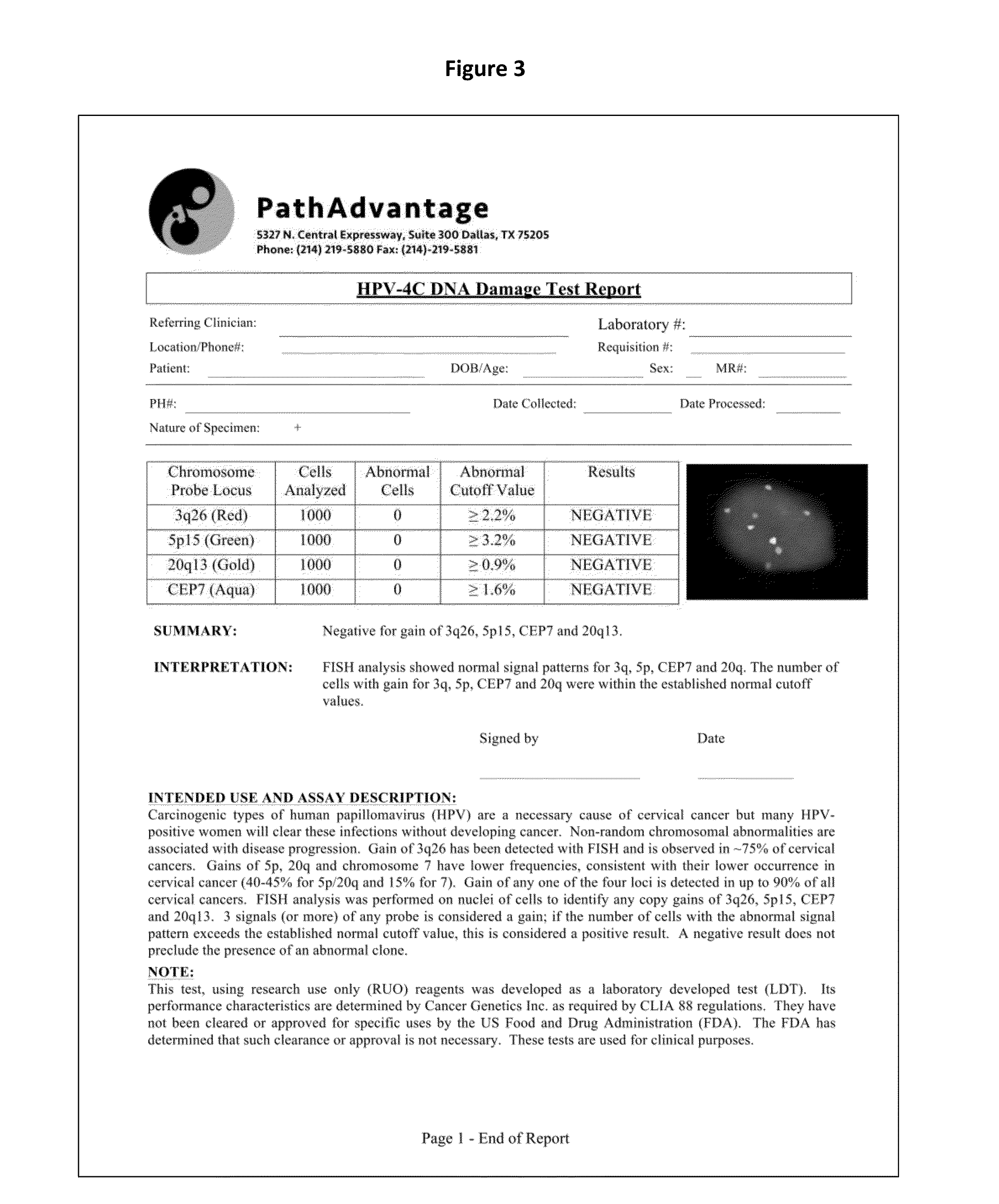
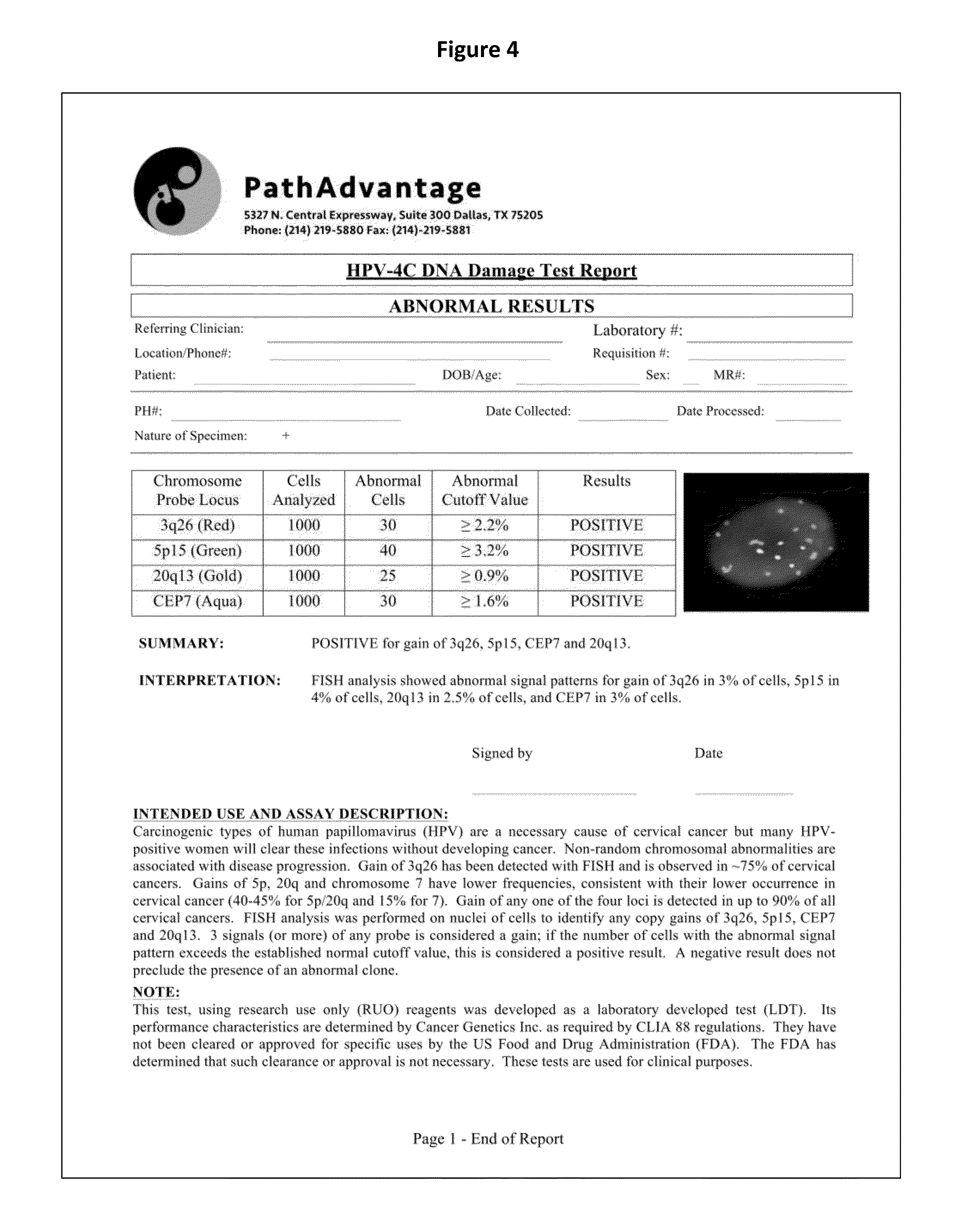
[0094](1) A method for identifying an abnormal sample of cells comprising:[0095]a) hybridizing a set of chromosomal probes to the sample, wherein the set comprises probes to 3q26, 5p15, CEP7, and 20q13;[0096]b) evaluating cells of the sample to detect and quantify the presence of each probe in the set;[0097]c) categorizing the evaluated cells of the sample as normal or abnormal, wherein the normal cells contain exactly two copies of each probe in the set and the abnormal cells do not contain exactly two copies of each probe in the set;[0098]d) calculating the percentage of the abnormal cells in the evaluated cells of the sample; and[0099]e) identifying the sample of cells as abnormal if the percentage of abnormal cells in the evaluated cells is greater than or equal to a cut-off value of 0.3%.
(2) The method of (1), wherein the sample of cells is a sample of cervical, vaginal, or anal cells.
(3) The method of (2), wherein the abnormal cells are selected from the group consisting of: c...
example 1
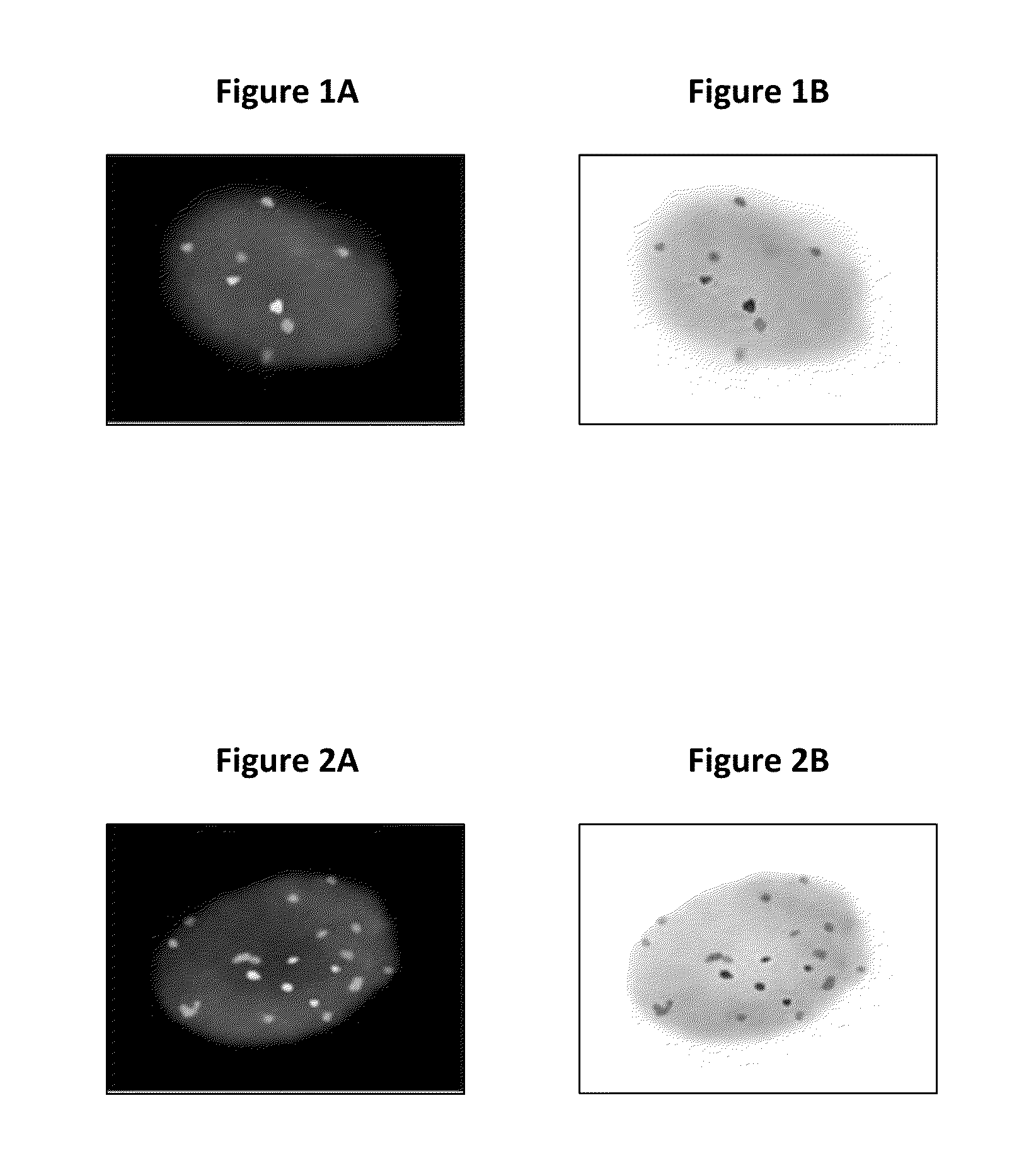
HPV 4C FISH Assay
[0177]a. Reagent Preparation
[0178]20×SSC: Powered 20×SSC (264 g) was mixed with 900 ml DI water using a magnetic stir plate and stir bar. The pH was adjusted to 7.0-7.5 with HCl. The total volume brought up to 1000 ml. The solution was filtered through a 0.45 μm pore filtration unit into the collection / storage bottle. This solution could be stored at room temperature for up to 6 months.
[0179]2×SSC: A volume of 20×SSC (100 ml) was mixed with 900 ml DI water. The solution was filtered through a 0.45 μm pore filtration unit into the collection / storage bottle. This solution could be stored at room temperature for up to 6 months. Any used solution was discarded at the end of the day.
[0180]2×SSC / 0.1% NP-40: A volume of 20×SSC (100 ml) was mixed with 899 ml DI water and 1 ml of NP-40. The pH was adjusted to about 7.0 (+ / −0.2). The solution was filtered through a 0.45 μm pore filtration unit into the collection / storage bottle. This solution could be stored at room temperatu...
example 2
HPV 4C FISH—Manual Scoring
[0205]Slides were prepared from cervical or vaginal ThinPrep Pap Test specimen according to the preparation and hybridization protocol discussed in Example 1 and were stored at −20° C. until they were ready to be analyzed with the following procedure.
[0206]a. Slide Analysis
[0207]Probe signals and DAPI counterstain were visualized using the following fluorescent filters:[0208]1. DAPI single bandpass (360 nm excitation, 460 nm emission): for viewing nuclei—in Filter Wheel position 1[0209]2. Green single bandpass (496 nm excitation, 520 nm emission): for viewing 5p15 (D5S2095)—in Filter Wheel position 3[0210]3. Red single bandpass (593 nm excitation, 612 nm emission): for viewing 3q26 (TERC)—in Filter Wheel position 2[0211]4. Aqua single bandpass (431 nm excitation, 480 nm emission): for viewing chromosome 7 (Cen7)—in Filter Wheel position 4[0212]5. Gold single bandpass (525 nm excitation, 551 nm emission): for viewing 20q13 (D20S911)—in Filter Wheel position ...
PUM
| Property | Measurement | Unit |
|---|---|---|
| temperature | aaaaa | aaaaa |
| volume | aaaaa | aaaaa |
| volume | aaaaa | aaaaa |
Abstract
Description
Claims
Application Information