
Skin substitutes and methods for hair follicle neogenesis

a technology of hair follicles and skin substitutes, applied in the field of skin substitutes and methods for hair follicle neogenesis, can solve the problems of limited patient use, skin substitutes that cannot perform all the functions of normal skin, and cells susceptible to loss through minor trauma and damage through ultraviolet ligh

Active Publication Date: 2016-06-30
THE HENRY M JACKSON FOUND FOR THE ADVANCEMENT OF MILITARY MEDICINE INC
View PDF2 Cites 6 Cited by
  • Summary
  • Abstract
  • Description
  • Claims
  • Application Information

AI Technical Summary

Benefits of technology

The patent text describes a new method for creating human hair follicles using a skin substitute made of neural crest-derived mesenchymal cells and epithelial cells. These cells are able to induce the formation of morphologically-correct and fully-developed hair follicles that can be used for treating skin injuries and conditions where hair follicle formation is needed. The method involves combining these cells in a specific way to create a skin substitute that can be used to induce hair follicle formation. The resulting skin substitute is a promising tool for restoring normal skin function and appearance.

Problems solved by technology

However, currently available skin substitutes cannot perform all the functions of normal skin.
For example, hair follicle (HF) neogenesis is not observed using any currently available skin substitute, which limits their use in patients.
In addition, any stem cells that might exist in skin lacking HFs are located in superficial layers of the epidermis, making the cells susceptible to loss through minor trauma and damage through ultraviolet light.
DP cells from rodents induce HFs in a variety of assays (reviewed in Ohyama et al., 2009), but it has been difficult to grow human DP cells that maintain inductive capacity in culture (Ohyama et al., 2013).
This is a significant problem since DP cells must be enriched in culture to expand the cells needed for successful clinical use.
However, while chimeric HFs are highly valuable as investigative tools, they lack clinical utility because the HFs produced by these methods are not fully human constructs (but instead are chimeric rodent / human constructs), are not completely developed, contain hair shafts in the wrong anatomical location, do not exhibit long-term graft survival and normal HF cycling, and / or do not form HFs that contain sebaceous glands.
In addition, HFs produced by such methods tend to grow in variable and uncontrollable directions, resulting in unnatural looking hair.
Thus, the follicles produced by such methods are not useful for human HF neogenesis in skin lacking hair follicles.

Method used


Image

  • Skin substitutes and methods for hair follicle neogenesis
  • Skin substitutes and methods for hair follicle neogenesis
  • Skin substitutes and methods for hair follicle neogenesis

Examples


example 1

[0163]Human DP cells isolated from temporal scalp dermis (Promocell, Heidelberg, Germany) from one male and five female donors were propagated in vitro according to methods described below. Alkaline phosphatase activity, a DP marker which correlates with hair-inducing capacity (Ohyama et al., 2013), was measured in vitro using the BCIP / NBT substrate (Sigma-Aldrich, St. Louis, Mo.) on passage 5 DP cells. To screen for trichogenicity, the reconstitution described in Zheng et al., 2005, and Kang et al., 2012 was used with minor modifications.

[0164]Human DP cells induced HFs when grown as spheroids, but not as monolayers, when coinjected with mouse epidermal aggregates in a reconstitution assay (Table 1), similar to the results of others (Kang et al, 2012). Skin substitutes (of which DECs are one type) constructed with human DP cells and human neonatal foreskin keratinocytes (NFKs) were grafted onto female nude mice. Eight weeks after grafting, HFs were observed in mice grafted with hum...

example 2

[0176]Studies were done using human dermal papilla cells grown from the temporal scalp of 6 individuals (HDP47, HDP44, HDP41, HDP43, HDP60, and HDP52) and human dermal papilla cells grown from the occipital scalp from 7 individuals (1-7). All cells were combined with human foreskin keratinocytes and rat collagen type I as described as in Example 1. The alkaline phosphatase activity of cells was measured in monolayer cultures before grafting.

[0177]Table 2 shows the results of the grafting experiments comparing human dermal papilla cells isolated from the temporal versus occipital scalp.

[0178]The second column in Table 2 shows the number of grafts in which the epidermis showed a typical human epidermal morphology by H&E staining. Dermal papilla cells from the temporal scalp supported the development of the human keratinocytes into a stratified squamous epithelium much better than those from the occipital scalp. In many grafts with occipital dermal papilla cells, the mouse keratinocyte...

example 3

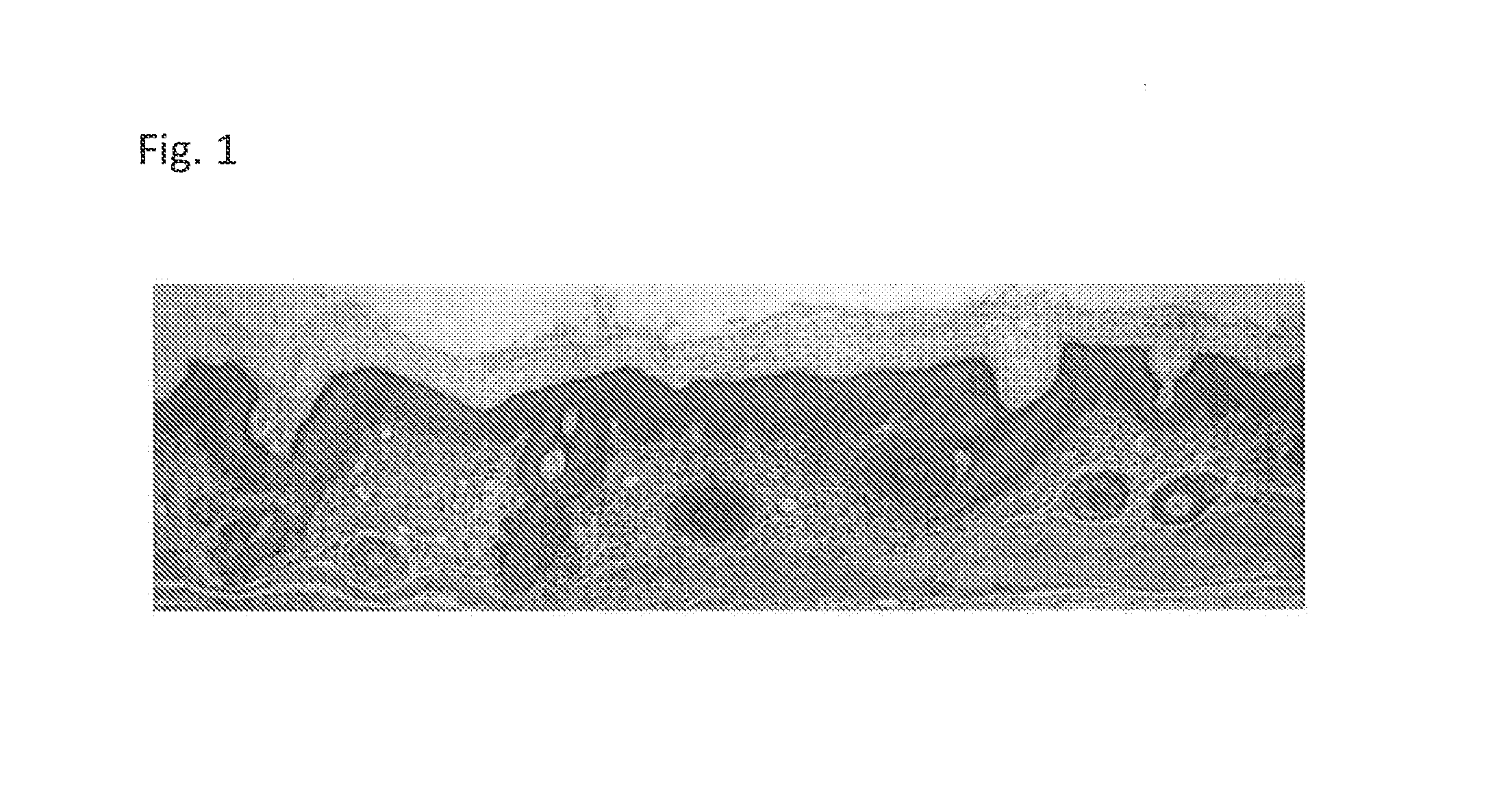
[0181]Grafted dermal-epidermal composites constructed from human dermal and epidermal cells (neural crest-derived human dermal papilla cells from temporal scalp dermis and neonatal foreskin epidermal cells) were evaluated 8, 12, and 15 weeks after grafting into nude mice. FIG. 13 provides H&E stain showing a hair shaft emerging from the infundibulum of a human hair follicle in a construct 8 weeks after grafting. FIG. 14 is a dermal-epidermal composite photographed under a light microscope after 10 weeks. Hair shafts can be seen emerging from the grafted region. FIG. 15A provides a representative graft with hair shafts visible 12 weeks after grafting, while FIG. 15B provides a magnified view showing the presence of pigmented hair shafts.

[0182]FIGS. 16A-C show that different hair follicle stages could be detected in grafts after 15 weeks. The grafts containing dermal papilla cells grown in monolayers, and those grown as spheroids, had telogen hair follicles, confirmed by club-like app...


PUM

PropertyMeasurementUnit
diameteraaaaaaaaaa
diameteraaaaaaaaaa
temperatureaaaaaaaaaa

Abstract

This invention provides compositions in the form of skin substitutes comprising epithelial cells and mesenchymal cells, wherein the mesenchymal cells are not isolated from the occipital or nape region of the scalp, as well as methods for using the same.

Description

CROSS-REFERENCE TO RELATED APPLICATIONS[0001]This application is the United States national stage entry under 35 U.S.C. §371 of International Application No. PCT / US2014 / 036351, filed May 1, 2014 and published as WO 2014 / 179559 A1, which claims priority to U.S. Provisional Application No. 61 / 819,332, filed May 3, 2013. The contents of these applications are each incorporated herein by reference in their entirety.STATEMENT OF GOVERNMENT INTEREST[0002]This invention was made in part with support from the U.S. Government. Accordingly, the Government has certain rights in this invention.FIELD OF THE INVENTION[0003]The present invention relates to compositions in the form of skin substitutes and microspheres comprising neural crest-derived mesencymal cells, such as hair follicle dermal cells, e.g., dermal papilla (“DP”) cells, or dermal sheath cells, that are capable of inducing hair follicles (“HFs”). The present invention also relates to methods, uses, and compositions for inducing HF g...

Claims


Application Information

Patent Timeline
Patent Type & Authority Applications(United States)
IPC IPC(8): A61L27/38A61L27/24
CPCA61L27/3886A61L27/24A61L2430/18A61L27/3813A61L27/3804A61L27/60A61P17/00A61P17/02A61P17/14
Inventor THANGAPAZHAM, RAJESHDARLING, THOMAS N.LI, SHAOWEI
Owner THE HENRY M JACKSON FOUND FOR THE ADVANCEMENT OF MILITARY MEDICINE INC
Learn More