Sheathed Duodenoscope

a duodenoscope and endoscope technology, applied in the field of medical devices, can solve the problems of inability to apply autoclave, inconvenient use, and inability to disinfect duodenoscopes, and achieve the effect of preventing interference from stray light and preventing stray light interferen

Active Publication Date: 2016-08-11
SHENYANG SHENDA ENDOSCOPE
View PDF6 Cites 20 Cited by
  • Summary
  • Abstract
  • Description
  • Claims
  • Application Information

AI Technical Summary

Benefits of technology

[0011]1. Design to prevent stray light interference, wherein there is a capsule containing transparent fluid, which will become broken under a cooperative action of the end cap and the distal end portion of the shaft of the endoscope, and the transparent liquid contained in the capsule filling a space between the end cap and the distal end portion of the shaft of the endoscope, preventing interference from stray light.
[0012]2. All contacting surfaces between endoscope and patient are covered by disposable component, to protect endoscope from cross contamination.

Problems solved by technology

However, inadequate disinfection of duodenoscopes has been a top medical hazards.
Autoclave cannot be applied due to the intolerance of the charge-coupled device CCD—a component material of endoscope, to high temperature and pressure.
In the operation part, steel wire was used to control the forceps elevator to lift up and down, and the structure is quite complicated with wire leading orifices.
The disinfection is inadequate due to disinfection limitations.
This patented duodenoscope has even more complicated structure inside and carries more risks than common duodenoscope.
However, it resulted in compromised image quality due to that stray light is produced as there are gaps between the distal end of endoscope and covering end cap.

Method used


Image

  • Sheathed Duodenoscope
  • Sheathed Duodenoscope
  • Sheathed Duodenoscope

Examples


Embodiment Construction

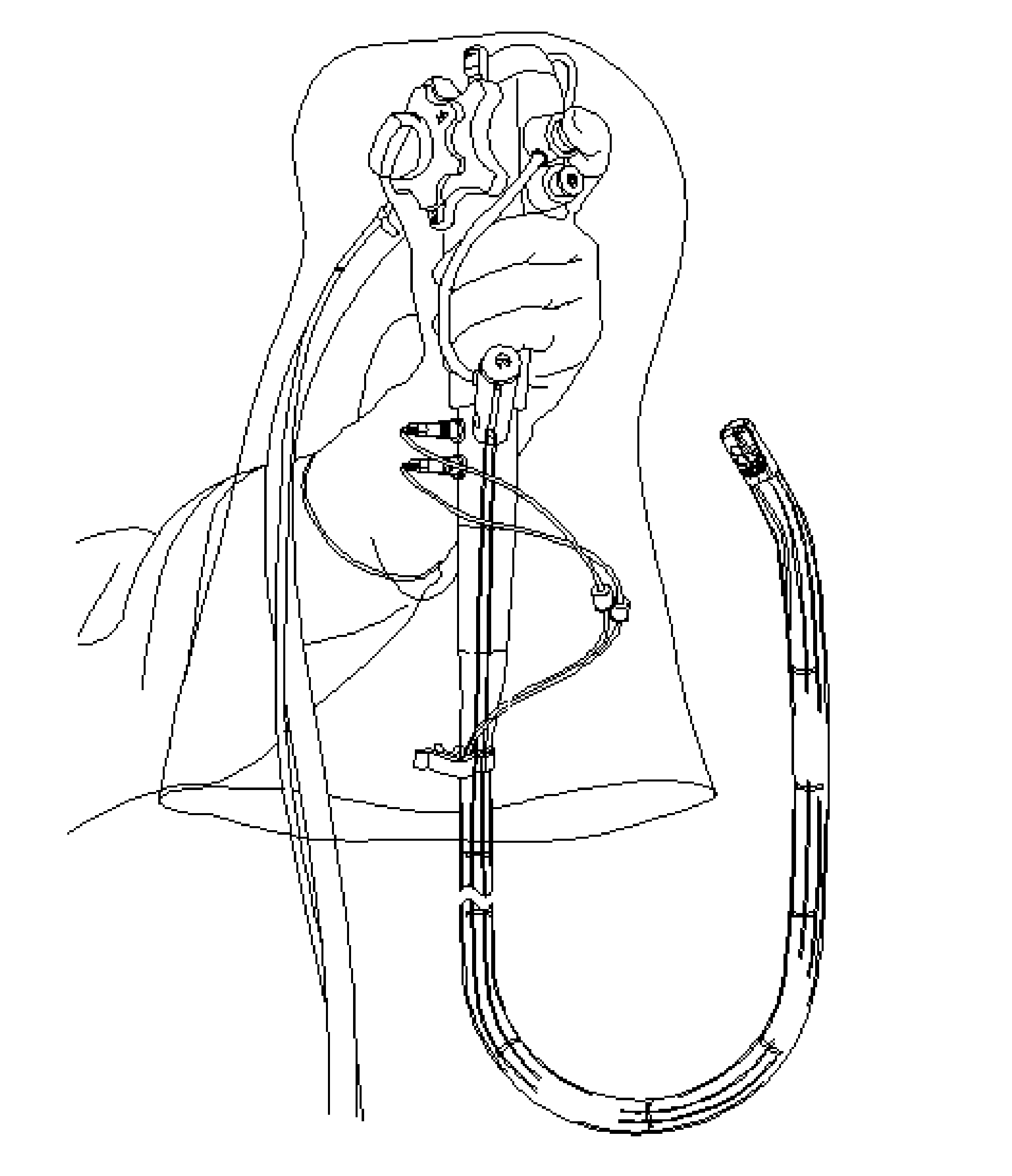
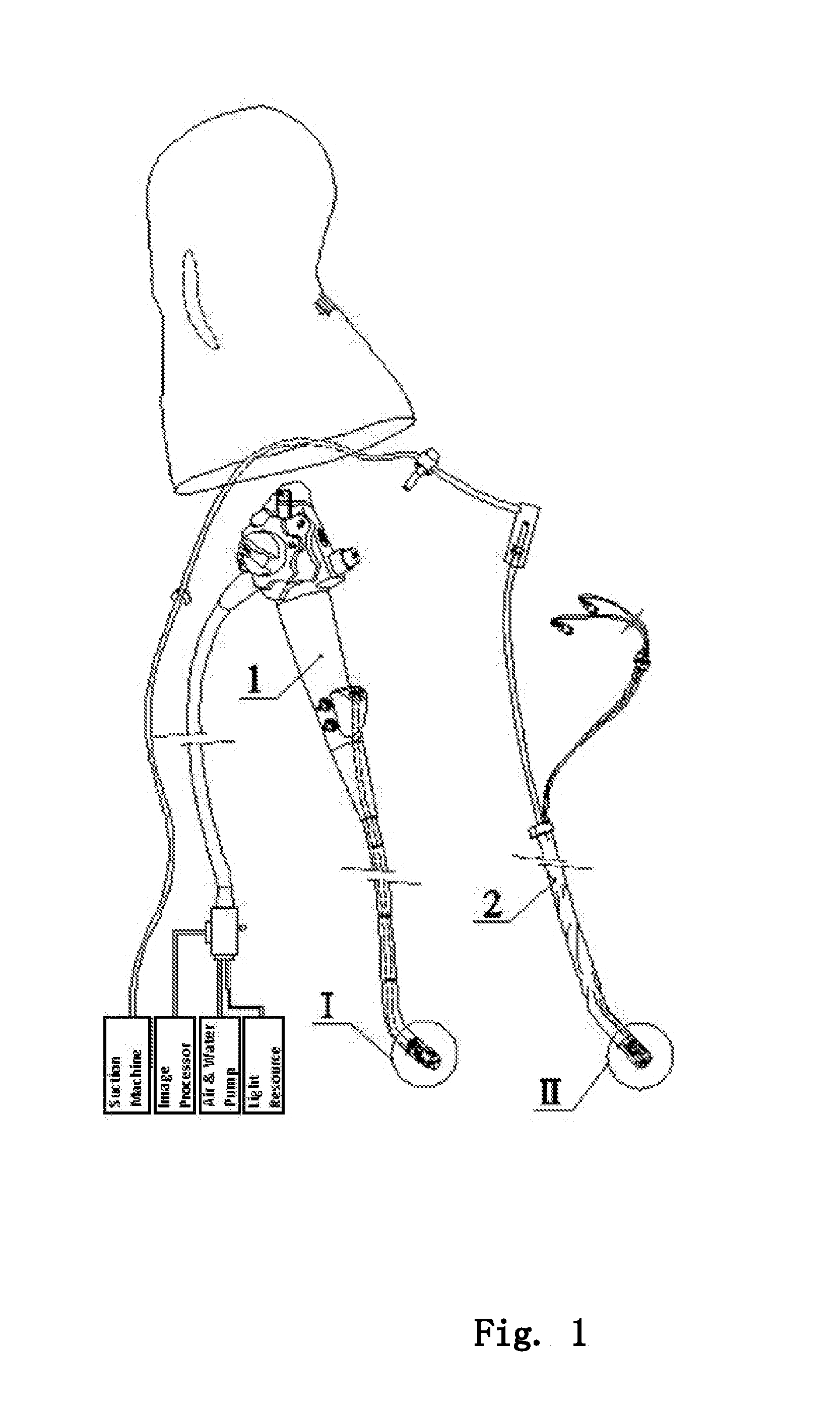
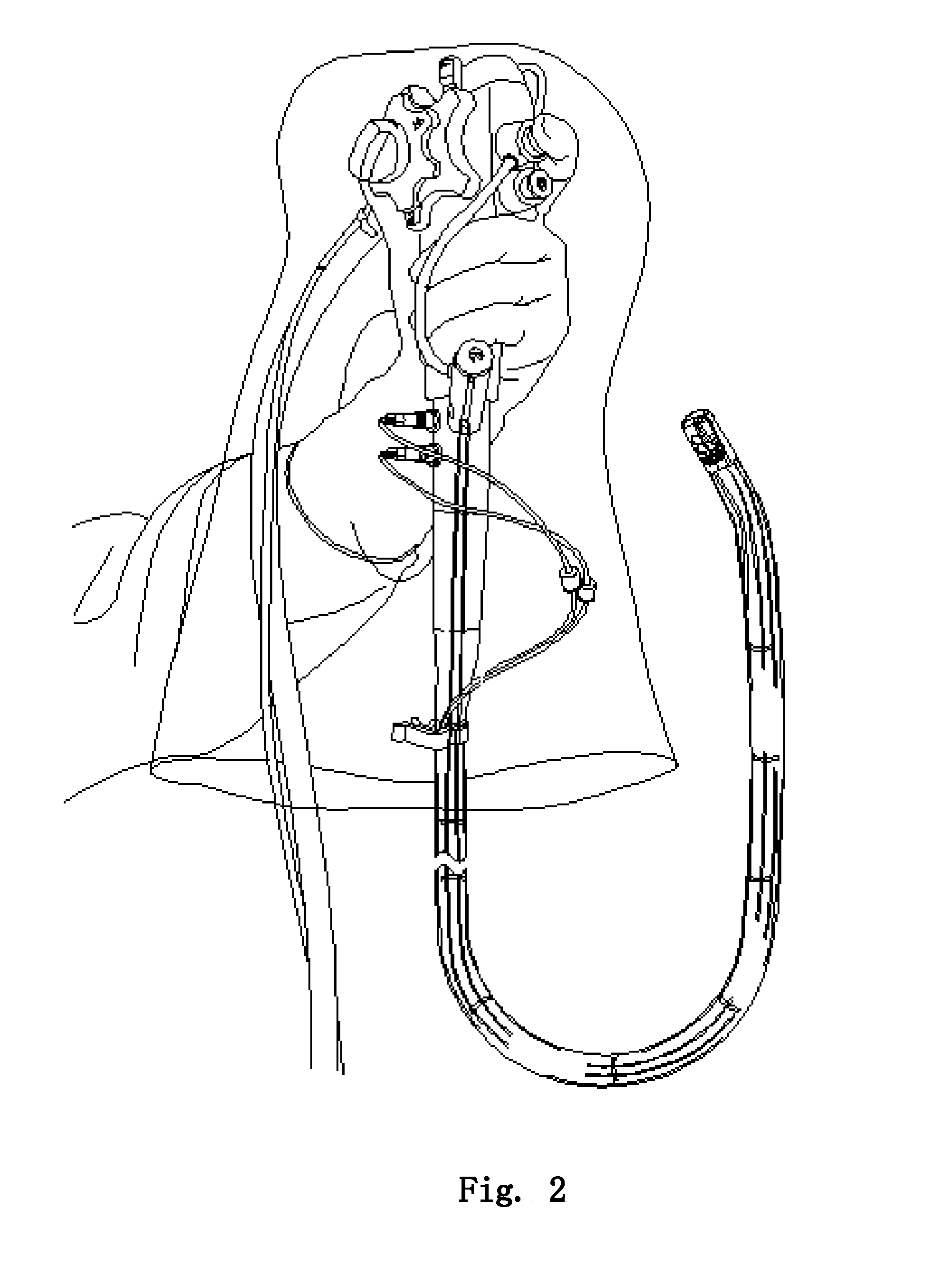
[0027]As shown in FIG. 1-13, this invention comprises two parts: one is modified endoscope 1, as shown in FIG. 3, wherein there is an elongated slot on the distal end portion 1.1 of the shaft of the endoscope with a forceps elevator 1.2 disposed inside; another part is the disposable component 2, as shown in FIG. 4-13, which covers the endoscope to protect it from being contaminated, including both the cuff 2.4 and the end cap 2.1 covering the out surface, as shown in FIG. 11-12, the disposable biopsy channel 2.6 connecting with the end cap, covering totally the endoscope 1 on both inside and outside. There is the capsule 2.2 filled with transparent liquid being provided in between the end cap 2.1 and the distal end portion 1.1 of the shaft of the endoscope, which will become broken under a cooperative action of the end cap 2.1 and the distal end portion 1.1 of the shaft of the endoscope. After the capsule 2.2 being broken, the transparent liquid contained in the capsule 2.2 will fi...


PUM


Abstract

A sheathed endoscope, includes: an endoscope and a disposable component. The disposable compartment protects the inner and outer surfaces of the endoscope. The disposable compartment includes: a cap of endoscope tip in the distal end of endoscope, a disposable tube for biopsy forceps, and an outside sheath capsule for endoscope shaft. The tip cap connects the sheath capsule and the disposable tube for biopsy forceps. There are multiple windows set on the lateral side of endoscope tip, wherein pills containing transparent fluid are placed somewhere in between of the tip cap and the distal end of endoscope. The pills will be crushed by co-fraction of the tip cap and the distal end of endoscope, to release the transparent fluid to fill in the space formed between the tip cap and the distal end of the endoscope. This disposable sheath compartment in the invention will cover all surfaces of the endoscope with disposable material, preventing contaminations on endoscope during endoscopy, meanwhile there is anti-glaring design, to prevent producing of glaring in the space formed between the tip cap and the distal end of endoscope.

Description

FIELD OF THE INVENTION[0001]An invention in the field of medical device, to be specified, a sheathed endoscope.BACKGROUND OF THE INVENTION[0002]Duodenoscope is widely used in the diagnosis and treatments of diseases in liver, gall bladder and pancreas, especially as an essential and irreplaceable device for the procedure of Endoscopic retrograde cholangiopancreatography ERCP and minimally invasive surgery. However, inadequate disinfection of duodenoscopes has been a top medical hazards. Autoclave cannot be applied due to the intolerance of the charge-coupled device CCD—a component material of endoscope, to high temperature and pressure. In the operation part, steel wire was used to control the forceps elevator to lift up and down, and the structure is quite complicated with wire leading orifices. The disinfection is inadequate due to disinfection limitations. This patented duodenoscope has even more complicated structure inside and carries more risks than common duodenoscope.[0003]T...

Claims


Application Information

Patent Timeline
Patent Type & Authority Applications(United States)
IPC IPC(8): A61B1/00A61B1/273A61B1/018
CPCA61B1/00142A61B1/2736A61B1/018A61B1/00137A61B1/00096A61B1/00098A61B1/00101A61B1/00135A61B1/00177A61B1/0615
Inventor JIANG, KERANGWU, XIANGYANGZHOU, ZHENYUCHEN, CHONGZHANG, JIANXU, MING
Owner SHENYANG SHENDA ENDOSCOPE
Why Eureka
Try Eureka