
Methods for detecting, monitoring and treating lymphedema

a lymphedema and lymphedema technology, applied in the field of lymphedema detection, monitoring and treating, can solve the problems of long-term suffering, inability to completely reverse the lymphedema therapy, and associated medical costs, and achieve the effect of enhancing patient access to lymphedema detection, monitoring and treatment, and simplifying body image acquisition and processing

Inactive Publication Date: 2016-08-18
LYMPHATECH INC
View PDF4 Cites 25 Cited by
  • Summary
  • Abstract
  • Description
  • Claims
  • Application Information

AI Technical Summary

Benefits of technology

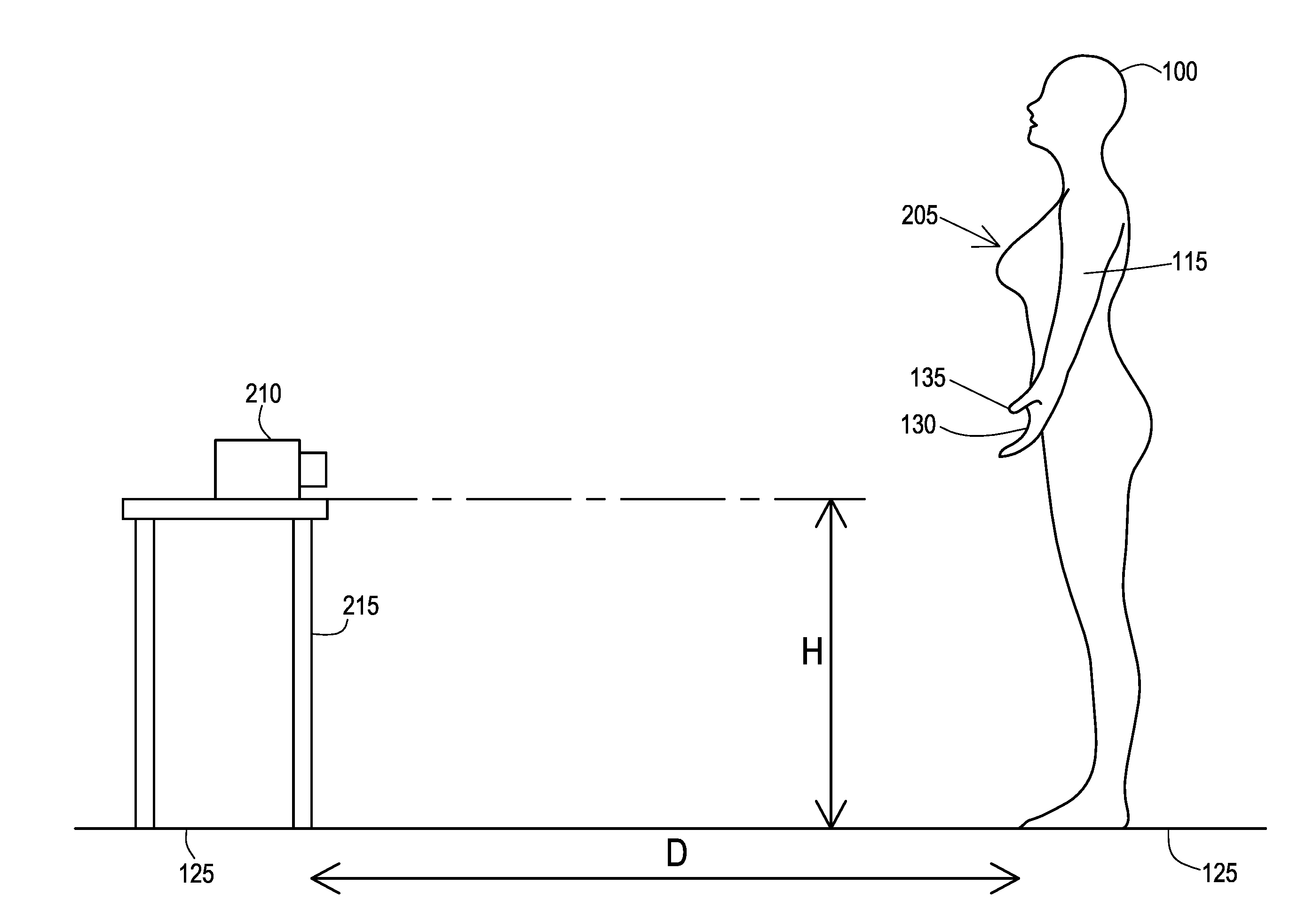
The present invention is about using a special imaging device called a ToF to detect, monitor, and treat lymphedema. This allows for easier and more accessible detection and treatment of the condition, as well as fitting compression garments. The method can be used at home or other non-clinical settings. The invention also allows for range of motion assessment and patient motion tracking. Overall, it improves the ability to care for patients with lymphedema and enhances the patient's quality of life.

Problems solved by technology

Even in early stage breast cancer, various studies have shown that when lymphedema progressed, lymphedema therapy could not completely reverse it.
Where lymphedema manifests, the patient is likely to suffer from lymphedema for the remainder of her life, thus leading to long-term suffering, as well as the attendant medical costs associated with the treatment of such a chronic condition.
Essentially anyone with a breast cancer diagnosis who undergoes surgery, chemotherapy and / or radiation therapy is at relative risk for generating lymphedema.
Surgery or treatment for prostate, colon and testicular cancers may result in lymphedema, particularly when lymph nodes have been removed or damaged.
However, while risk does diminish, some patients may be anatomically predisposed to developing lymphedema.
Excessive exposure to the sun can also lead to an inflammatory stimulus that overtaxes an already impaired lymphatic system, resulting in recurrent lymphedema.
Moreover, a significant number of lymphedema patients are obese or overweight and, therefore, would not be readily able to utilize off-the-shelf garments.
There is now a body of evidence demonstrating that waiting to treat BCRL when it becomes visible and symptomatic may not be optimal.
The diagnosis or early detection of lymphedema can be difficult.
Undiagnosed, or not treated effectively, lymphedema can progress into later stages of the condition resulting in a severe form of swelling called “elephantiasis.”
While tape measurement techniques are widely available to a broad scope of medical providers, the technique generally suffers from poor accuracy.
In short, tape measurement is not a very effective diagnostic tool because the current clinical threshold for a positive diagnosis of lymphedema is a 10% volume increase in a limb, while the tape measurement technique has been shown to exhibit as much as an about 8 to about 12% inaccuracy due to inter and intra-operator variability.
Water displacement can provide very accurate volumetric measurements, if performed correctly, but this technique is rarely implemented in a clinical setting due to challenges with implementation and hygiene concerns.
In short, few clinical settings are able to install and maintain a water tank of the size needed to accurately generate body part volume measurements or geometries needed to diagnose and treat lymphedema.
Volumetric measurement, when available, can allow for diagnosis of the onset of lymphedema in a patient, however, this measurement technique cannot be used to size and fit compression garments used to treat patients diagnosed with lymphedema.
However, the price of this device, believed to be about $30K, prevented widespread adoption of this device in the US.
Increased patient access to methods of detection is therefore paramount, even while patient access to clinicians can be restricted due to patient distance from clinicians, lack of necessary equipment in clinics and hospitals, and lack of available medical providers with training to use existing diagnosis techniques.

Method used


Image

  • Methods for detecting, monitoring and treating lymphedema
  • Methods for detecting, monitoring and treating lymphedema
  • Methods for detecting, monitoring and treating lymphedema

Examples


Embodiment Construction

[0031]Many aspects of the disclosure can be better understood with baseline to the Figures presented herewith. The Figures are intended to illustrate the various features of the present disclosure. Moreover, like references in the drawings designate corresponding parts among the several views. While several implementations may be described in connection with the included drawings, there is no intent to limit the disclosure to the implementations disclosed herein. To the contrary, the intent is to cover all alternatives, modifications, and equivalents.

[0032]The term “substantially” is meant to permit deviations from the descriptive term that do not negatively impact the intended purpose. All descriptive terms used herein are implicitly understood to be modified by the word “substantially,” even if the descriptive term is not explicitly modified by the word “substantially.”

[0033]The term “lymphedema” may include either primary or secondary lymphedema, the latter of which might also be...


PUM


Abstract

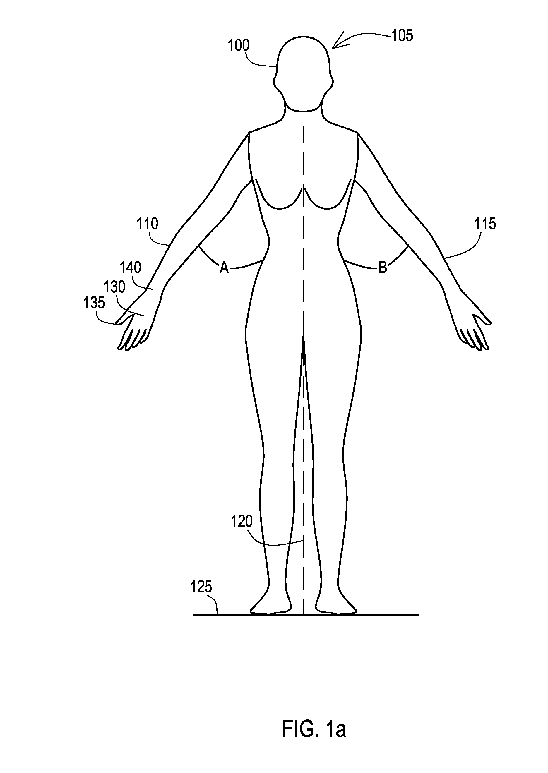
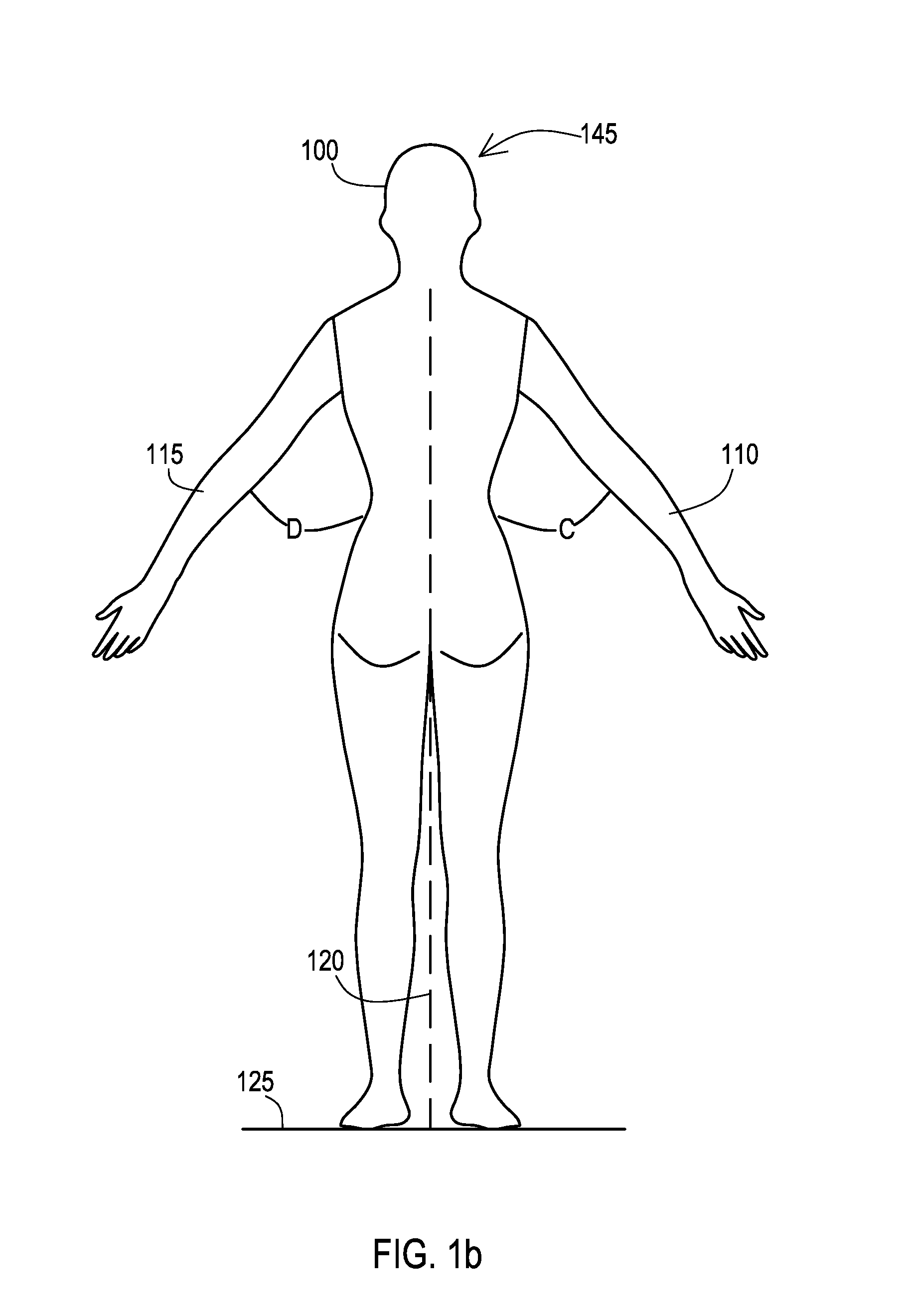
The present invention comprises methods for detecting, monitoring and treating lymphedema by using imaging devices to generate patient anatomical information from which body part volume measurements or geometries can be derived. Yet further, the present invention comprises methods of fitting compression garments for treatment of lymphedema by using the anatomical measurements derived from the patient anatomical information.

Description

CROSS-REFERENCE TO RELATED APPLICATIONS[0001]This application claims priority to U.S. Provisional Application No. 62 / 115,374 filed Feb. 12, 2015. This provisional application is incorporated herein in its entirety by this reference.FIELD OF THE INVENTION[0002]The present invention comprises methods for detecting, monitoring and treating lymphedema by using imaging devices to generate patient anatomical information from which body part volume measurements or geometries can be derived. Yet further, the present invention comprises methods of fitting compression garments for treatment of lymphedema by using the anatomical measurements derived from the patient anatomical information.BACKGROUND OF THE INVENTION[0003]Lymphedema can occur after any cancer or treatment that affects the flow of lymph through the lymphatic vessels, such as removal of lymph nodes. It may develop within days or many years after treatment. Most lymphedema develops within three years of surgery.[0004]Breast cancer...

Claims


Application Information

Patent Timeline
Patent Type & Authority Applications(United States)
IPC IPC(8): A61B5/00A61B5/107
CPCA61B5/4312A61B5/1071A61B5/0091A61B5/0077A61B5/1073A61B5/1121A61B5/41A61B5/4878
Inventor WEILER, MICHAEL J.DIXON, JAMES B.POKUTTA, SEBASTIANZINK, DANIELKASSIS, TIMOTHY
Owner LYMPHATECH INC
Learn More