
Proteasome inhibition assay and methods of use

a proteasome and assay technology, applied in the field of proteasome inhibition assay and methods of use, can solve the problems of cell lysate, difficult to establish a proper therapeutic window, and difficult to optimize drug levels in individual patients, and achieve the effect of detecting the modulation of proteasome activity

Active Publication Date: 2016-09-08
BECKMAN COULTER INC +1
View PDF0 Cites 0 Cited by
  • Summary
  • Abstract
  • Description
  • Claims
  • Application Information

AI Technical Summary

Benefits of technology

The patent describes an assay for detecting and monitoring proteasome activity in target cells, such as hematopoietic cells or neoplastic cells. The assay involves activating target cells with an IκB degradation agonist, contacting them with a labeled binding agent that specifically binds IκB, and detecting the amount of binding agent associated with the target cells. The assay can be used to screen compounds or drugs for proteasome inhibitory activity and to monitor the treatment of a disease or disorder in a patient. The assay can also be used to detect modulation of proteasome activity in a patient by measuring the level of IκB or other proteins. The patent provides a kit for performing the assay, including reagents, instructions, and optional additional reagents and controls.

Problems solved by technology

Current proteasome inhibitors can exhibit significant toxicity, which varies significantly from patient to patient.
At present, there is no biomarker assay to monitor the differential impact of proteasome inhibitors on normal vs tumor cell populations, making it difficult to establish a proper therapeutic window, or optimize drug levels in individual patients.
These assays require a cell lysate because fluorogenic substrates penetrate intact cells poorly, and fluorescent product can easily diffuse out of the cell.
It is therefore difficult to differentiate between a small number of target cells containing high concentrations of proteasomes (e.g., cancer cells) and a large number of normal cells containing low concentrations proteasomes.
In addition, unless the target cells can first be purified, it is not possible to distinguish the impact of any inhibitor on the target cells as opposed to normal cell populations in a sample.
As a result, these cell free assays provide little to no information on the functional effectiveness of inhibitors inside a target cell population.
In addition, the dilution introduced by cell lysis may cause dissociation of proteasome inhibitors with faster off-rates, making accurate measurement problematic.

Method used


Image

  • Proteasome inhibition assay and methods of use
  • Proteasome inhibition assay and methods of use
  • Proteasome inhibition assay and methods of use

Examples


example 1

IκB as a Biomarker for Proteasome Activity in Peripheral Blood Cells

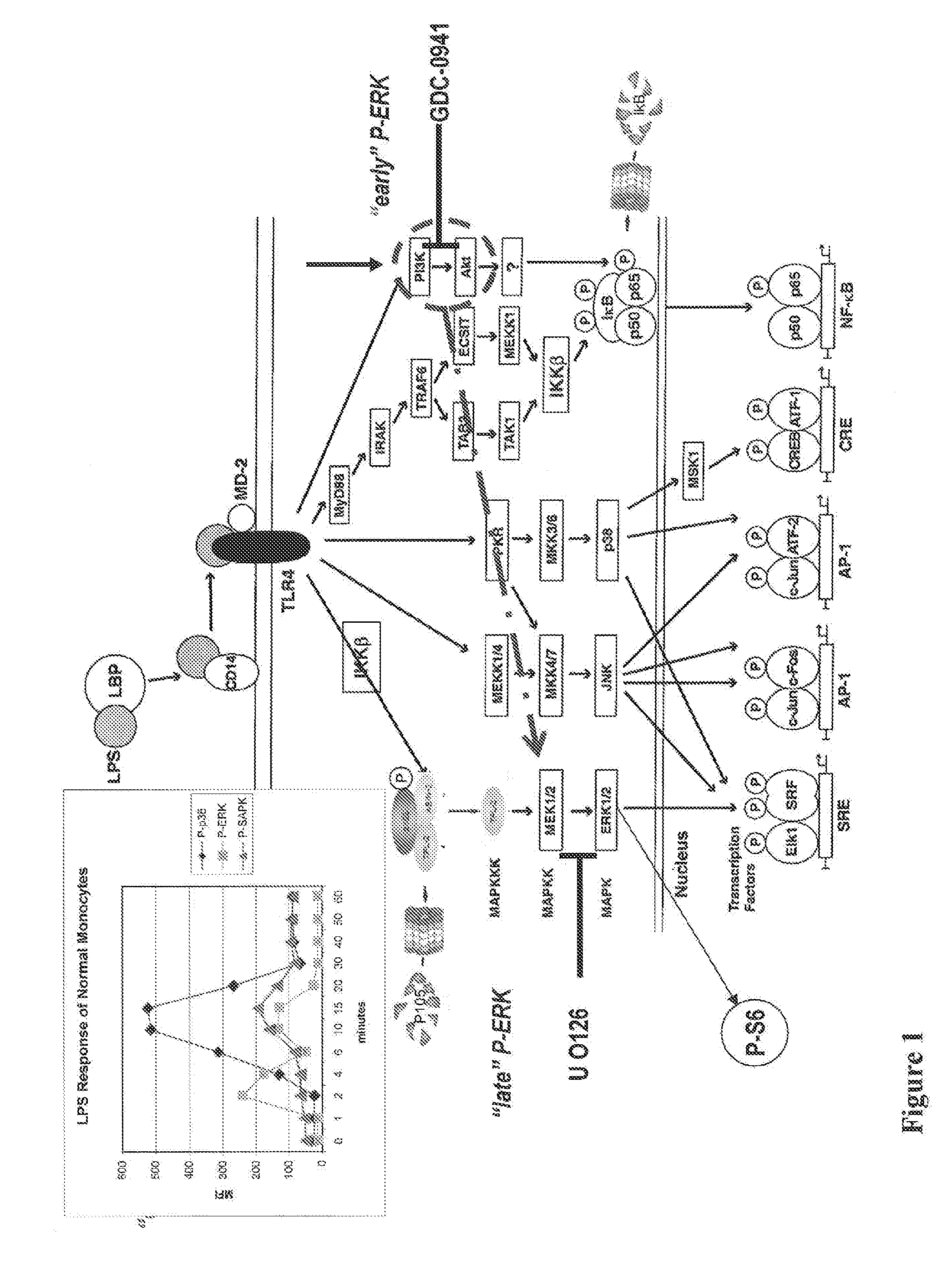
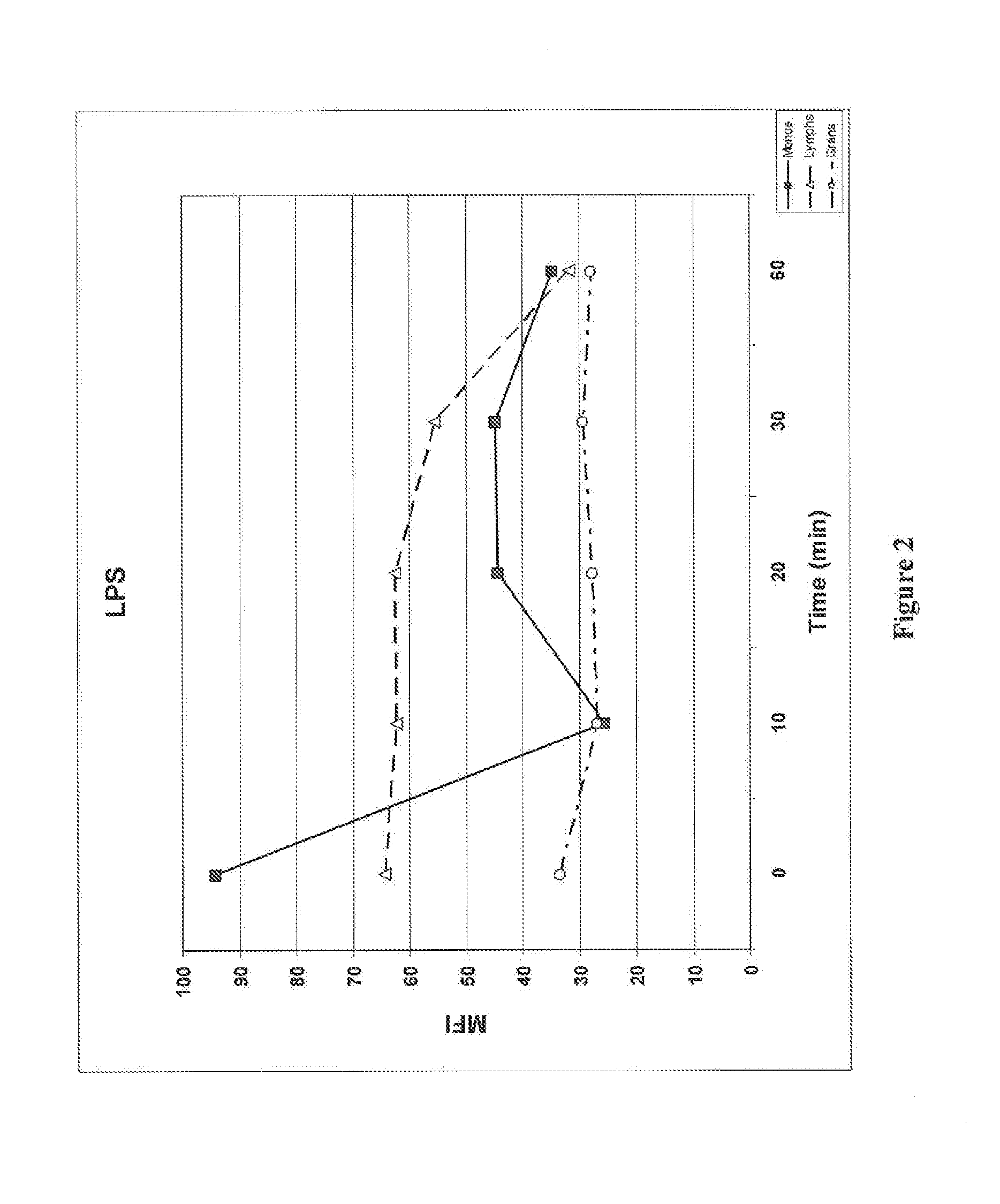
[0126]A flow-cytometry based assay to monitor proteasome activity in target cells has been developed. As shown in FIG. 1, exposure of cells (e.g., peripheral blood monocytes) to an activator of a signal transduction activator (e.g., LPS) results in phosphorylation of IκB via the TLR4 receptor. IκB normally sequesters the NF-κB complex in the cytoplasm and phosphorylation of IκB results in its ubiquination, tagging it for destruction by the proteasome. We have discovered that IκB levels can conveniently be used to monitor proteasome activity in peripheral bloods cells, providing for the development of a flow cytometry assay useful for monitoring and / or measuring the effect of drugs on proteasome activity.

Method

[0127]The assay generally comprises treating whole blood with an IκB degradation agonist and / or proteasome effector, fixing the blood cells, permeabilizing the fixed bloods cells, treating the fixed and permeab...

example 2

Monitoring of IκB and Phosphorylation of Signaling Pathways in Peripheral Blood WBCs Activated by LPS

[0135]Phosphorylation of signaling pathways activated by LPS, including ERK, Akt (activated by PI3 Kinase), and the ribosomal S6 protein (a marker of protein synthesis) were also monitored together with the degradation of IκB. The effect of MG-132, a proteasome inhibitor, and GDC-0941, a PI3 kinase inhibitor, were studied in monocytes. Whole blood samples were treated, activated, and processed as described in Example 1. The degradation of IκB in cells pretreated with MG-132 or GDC-0941 was monitored by flow cytometry as described in Example 1. The IκB degradation agonist used in this example was LPS. Whole blood cells were stained with antibodies to IκB, phosphorylated Akt (P-Akt: P-Ser473), phosphorylated ERK (P-ERK; P-Thr202 / P-Tyr204), and phosphorylated S6 (P-S6; P-Ser235 / P-Ser236). Monocytes were identified by antibodies to CD14 and lymphocytes were identified by antibodies to CD...

example 3

Monitoring of IκB and Phosphorylation of Signaling Pathways in Peripheral Blood WBCs Activated by TNF-α

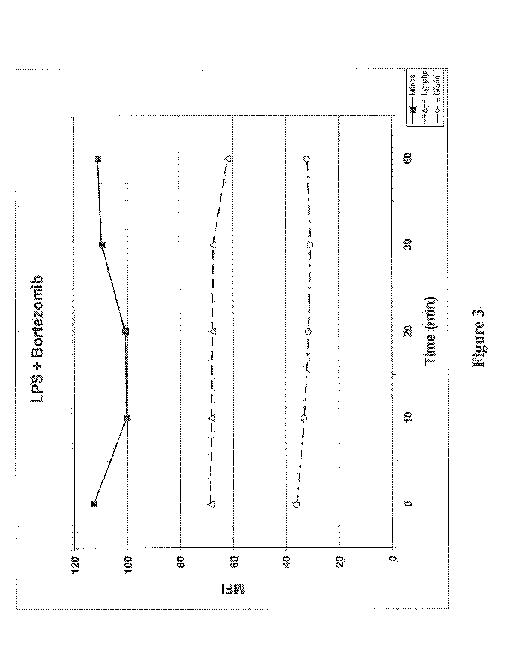
[0139]Phosphorylation of signaling pathways activated by TNF-α, including ERK, Akt, and the ribosomal S6 protein, was monitored together with the degradation of IκB in peripheral blood WBCs. Whole blood samples were treated, activated, and processed as described in Example 1 and the degradation of IκB in peripheral blood cells was monitored by flow cytometry as described in Example 1. The IκB degradation agonist used in this example was TNF-α. Whole blood cells were stained with antibodies to IκB, phosphorylated Akt (P-Akt: P-Ser473), phosphorylated ERK (P-ERK; P-Thr202 / P-Tyr204), and phosphorylated S6 (P-S6; P-Ser235 / P-Ser236). Monocytes were identified by antibodies to CD14 and lymphocytes were identified by antibodies to CD3 and CD19. A portion of the whole blood samples were treated with proteasome inhibitor Bortezomib before TNF-α activation as described in Example 1.

[0140]FIG...


PUM

PropertyMeasurementUnit
Covalent bondaaaaaaaaaa

Abstract

Assays that can measure the effect of proteasome inhibitors on target cells in a biological sample are provided. The assays include evaluation of the effects of proteasome inhibitors on proteasome activity in cells in a biological sample.

Description

CROSS-REFERENCE TO RELATED APPLICATION(S)[0001]This application is being filed on Oct. 3, 2014, as a PCT International Patent application and claims priority to U.S. Patent Application Ser. No. 61 / 892,778 filed on Oct. 18, 2013, the disclosure of which is incorporated herein by reference in its entirety.BACKGROUND OF THE INVENTION[0002]The proteasome is a protein complex that is present in all eukaryotic cells, and degrades intracellular proteins that are specifically marked for removal. As such, it regulates cell proliferation, apoptosis, and cell differentiation. In addition, it removes proteins that are misfolded, changed by aging, or damaged by oxidation. Empirical evidence suggests that cancer cells rely more heavily on intact proteasomal function, in part because they divide more frequently and in part because many of their regulatory pathways are disrupted due to aberrant protein structure.[0003]Inhibition of proteasome activity is therefore a therapuetic strategy for treatin...

Claims


Application Information

Patent Timeline
IPC IPC(8): G01N33/50G01N15/14
CPCG01N33/5041G01N33/5011G01N15/14G01N2015/008G01N2440/14G01N2015/149G01N2015/1006G01N2333/4704C12Q1/37G01N33/5047G01N33/582G01N33/4915C07K16/18G01N33/53C07K14/47G01N15/149G01N2015/016
Inventor SHANKEY, VINCENT T.GULNIK, SERGEILOPEZ, LIDICE L.HEDLEY, DAVID W.CHOW, SUE
Owner BECKMAN COULTER INC
Learn More