
Gpu-based system for performing 2d-3d deformable registration of a body organ using multiple 2d fluoroscopic views

a technology of 2d/3d fluoroscopic view and gpu, which is applied in the field of gpu-based system for performing 2d-3d rigid registration of body organs using multiple 2d fluoroscopic views, can solve the problems of high number of computations, inability to compute the pose from and inability to perform live 2d fluoroscopic images

Active Publication Date: 2016-09-08
BRONCUS MEDICAL
View PDF0 Cites 15 Cited by
  • Summary
  • Abstract
  • Description
  • Claims
  • Application Information

AI Technical Summary

Benefits of technology

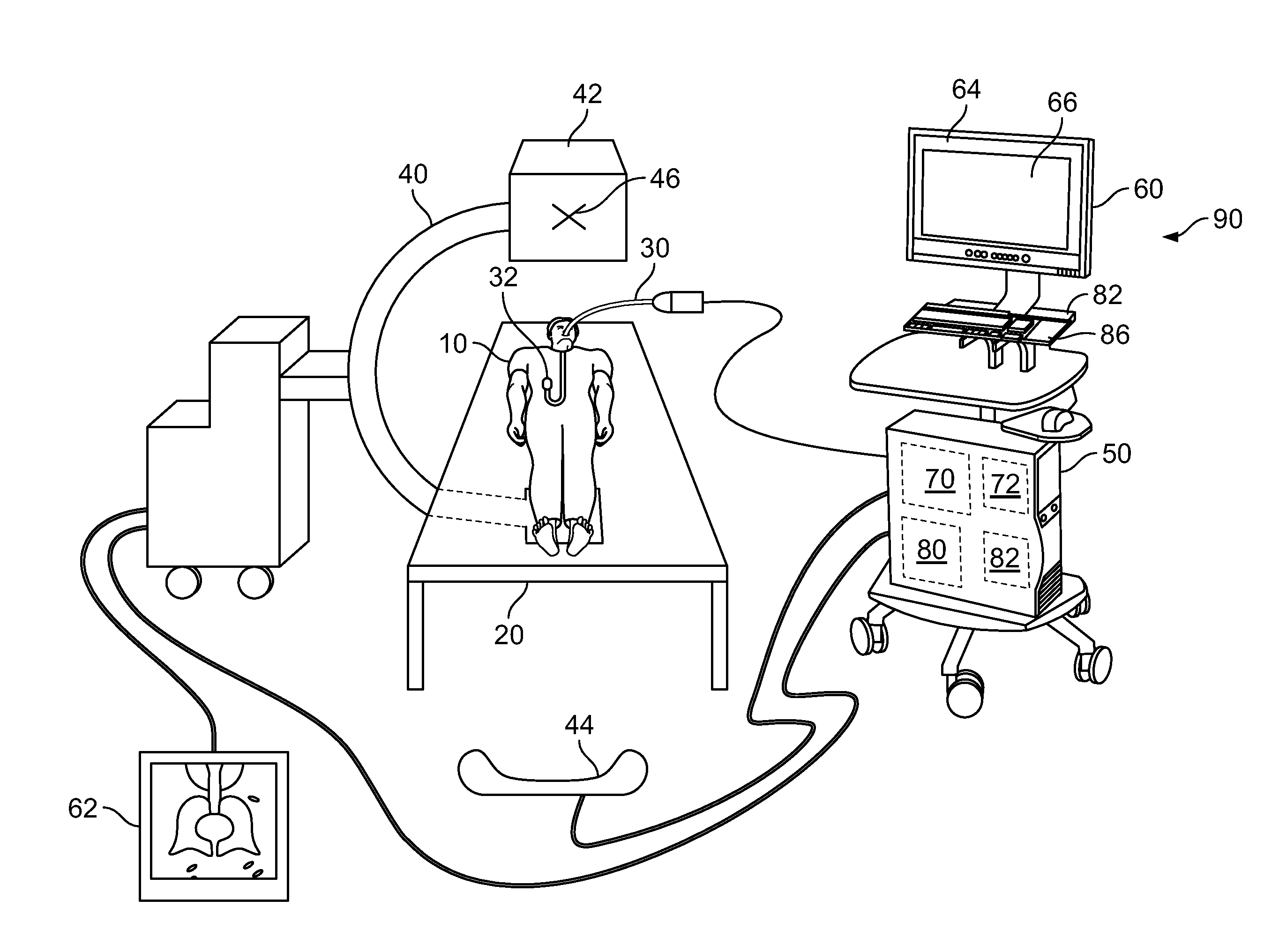
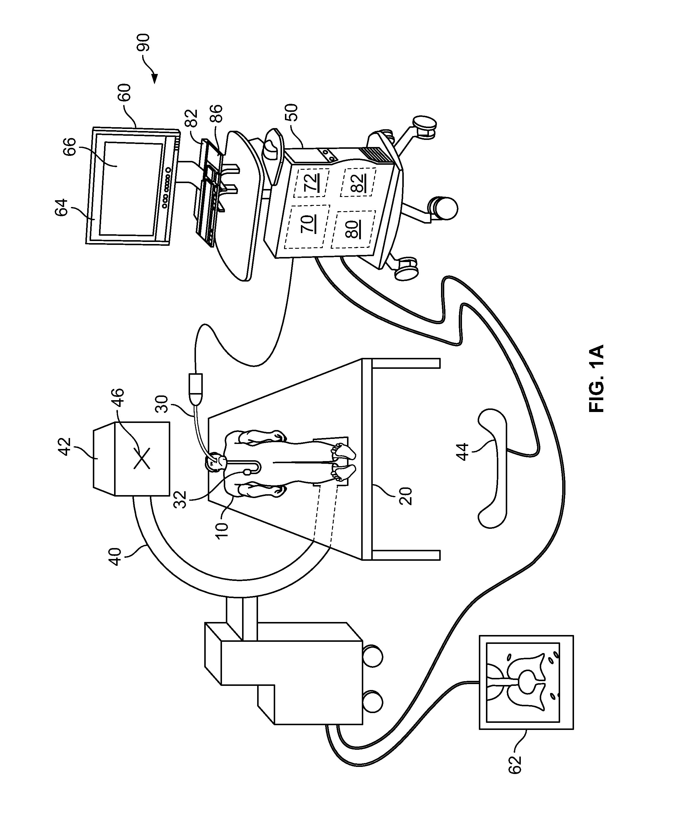
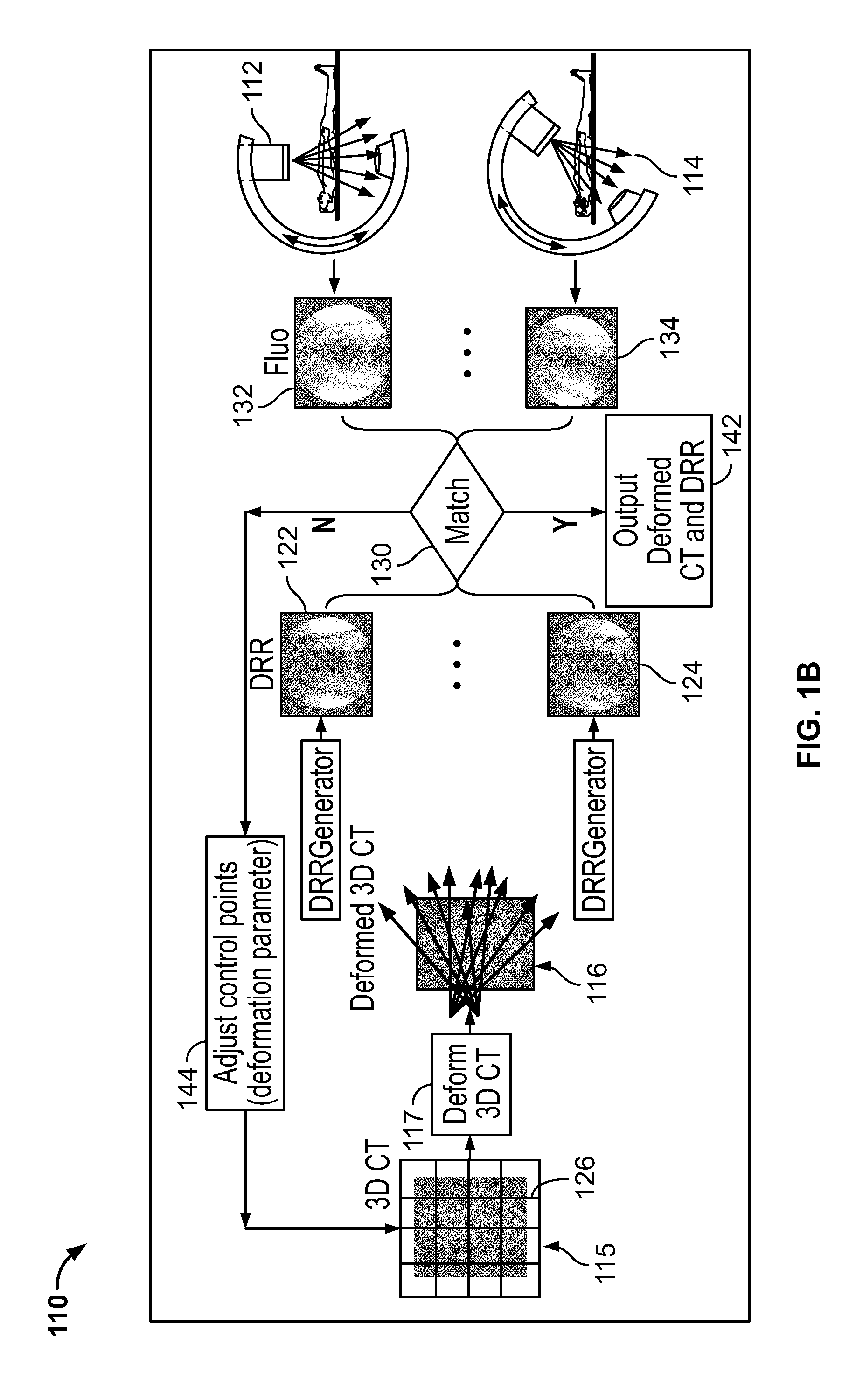
The present invention provides a method to fully recover a 3D deformation field, which means the deformation is recovered in 3D space and not just the 2D plane perpendicular to the projection direction. The method uses multiple 2D views "simultaneously" to determine a consistent 3D deformation field, which gives more accurate results. Computing the deformation field directly in 3D, the method can accurately handle motion of anatomical structures which are overlapping or occluded in 2D images. Recovering deformation in 3D space allows for a direct production of aDRR for navigation even if the camera position of the fluoroscopy unit is changed.

Problems solved by technology

Computing the pose from the live 2D fluoroscopic images, however, is not straightforward.
A challenge arises because the live 2D fluoroscopic images are missing information along the projection direction.
A challenge to performing 2D-3D rigid registration, however, is the high number of computations.
However, despite speeding up the registration, the accuracy established by the rigid 2D-3D rigid registration is reduced because of the patient's non-rigid motion between 3D image acquisition and 2D image acquisition.
In particular, the motion of the lung arising from the patient's repositioning, breathing, and beating heart introduces inaccuracies in the rigid registration.
This reduces the degrees of freedom and computation complexity, but undesirably results in two issues: (1) the “overlapping” anatomical structures in 2D image are deformed as one “layer” and therefore the deformation may not capture the “independent” 3D motion of the structures and (2) the 2D-2D deformation model does not allow for a previously occluded / out-of-camera-view structure to appear which may happen as part of an actual 3D deformation.
This reduces the accuracy of the 2D-2D deformation approach.
However, Rivest-Henault does not provide a solution to the more complicated task, namely, an alignment of the 2D-3D data for an entire or complete lung.
However, in the case of a GPU-based registration, parallelizing the DRR is not enough to speed up the process.
Parallelizing the DRR may only result in reducing computational time incrementally, not substantially for 2D-3D deformable registration.

Method used


Image

  • Gpu-based system for performing 2d-3d deformable registration of a body organ using multiple 2d fluoroscopic views
  • Gpu-based system for performing 2d-3d deformable registration of a body organ using multiple 2d fluoroscopic views
  • Gpu-based system for performing 2d-3d deformable registration of a body organ using multiple 2d fluoroscopic views

Examples


Embodiment Construction

[0055]Before the present invention is described in detail, it is to be understood that this invention is not limited to particular variations set forth herein as various changes or modifications may be made to the invention described and equivalents may be substituted without departing from the spirit and scope of the invention. As will be apparent to those of skill in the art upon reading this disclosure, each of the individual embodiments described and illustrated herein has discrete components and features which may be readily separated from or combined with the features of any of the other several embodiments without departing from the scope or spirit of the present invention. In addition, many modifications may be made to adapt a particular situation, material, composition of matter, process, process act(s) or step(s) to the objective(s), spirit or scope of the present invention. All such modifications are intended to be within the scope of the claims made herein.

[0056]Methods ...


PUM


Abstract

Systems and methods for assisting a physician in a medical intervention comprises performing a 2D-3D deformable registration, and more particularly, performing a 2D-3D registration based on multiple live 2D fluoroscopic views, and implemented on a multi-core processing framework such as a Graphics Processing Unit.

Description

CROSS-REFERENCE TO RELATED APPLICATIONS[0001]This application claims priority to provisional patent application number 62 / 128,885, filed Mar. 5, 2015, and entitled “GPU-BASED SYSTEM FOR PERFORMING 2D-3D DEFORMABLE REGISTRATION OF A BODY ORGAN USING MULTIPLE 2D FLUOROSCOPIC VIEWS.”BACKGROUND OF THE INVENTION[0002]Intra-operative poses of the lung of a patient may be computed from 2D live fluoroscopic images. The intra-operative poses can assist a physician in various medical procedures performed in the lung. For example, the pose information may be used to guide an instrument to a region of interest in the lung.[0003]Computing the pose from the live 2D fluoroscopic images, however, is not straightforward. A challenge arises because the live 2D fluoroscopic images are missing information along the projection direction. Hence, the 2D fluoroscopic images need to be aligned with a preoperatively acquired 3D CT to provide volumetric anatomical information, which leads to the 2D-3D rigid r...

Claims


Application Information

Patent Timeline
IPC IPC(8): G06T7/00G06T1/20
CPCG06T7/0024G06T1/20G06T2207/30196G06T2207/30061G06T2207/10064A61B6/032A61B6/0492A61B6/12A61B6/4441A61B6/4476A61B6/466A61B6/487A61B6/488A61B6/5205A61B6/5235A61B6/547A61B6/582G06T11/008G06T2207/10121G06T2207/10124G06T2211/428G06T7/30
Inventor LIU, YIXUNRAI, LAVWIBOWO, HENKY
Owner BRONCUS MEDICAL
Learn More