

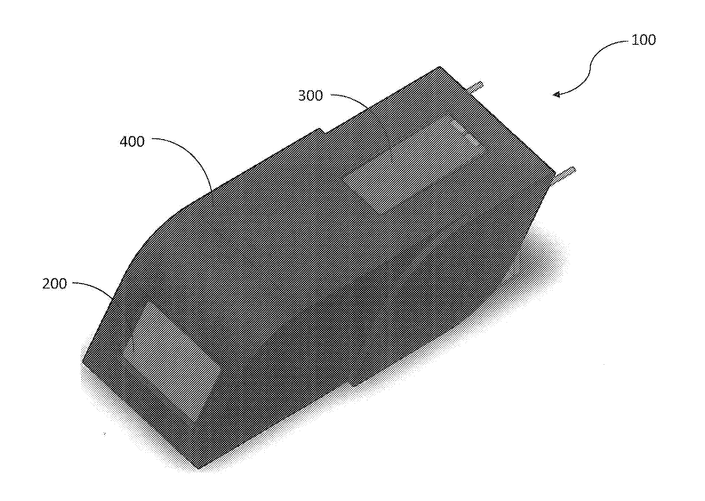
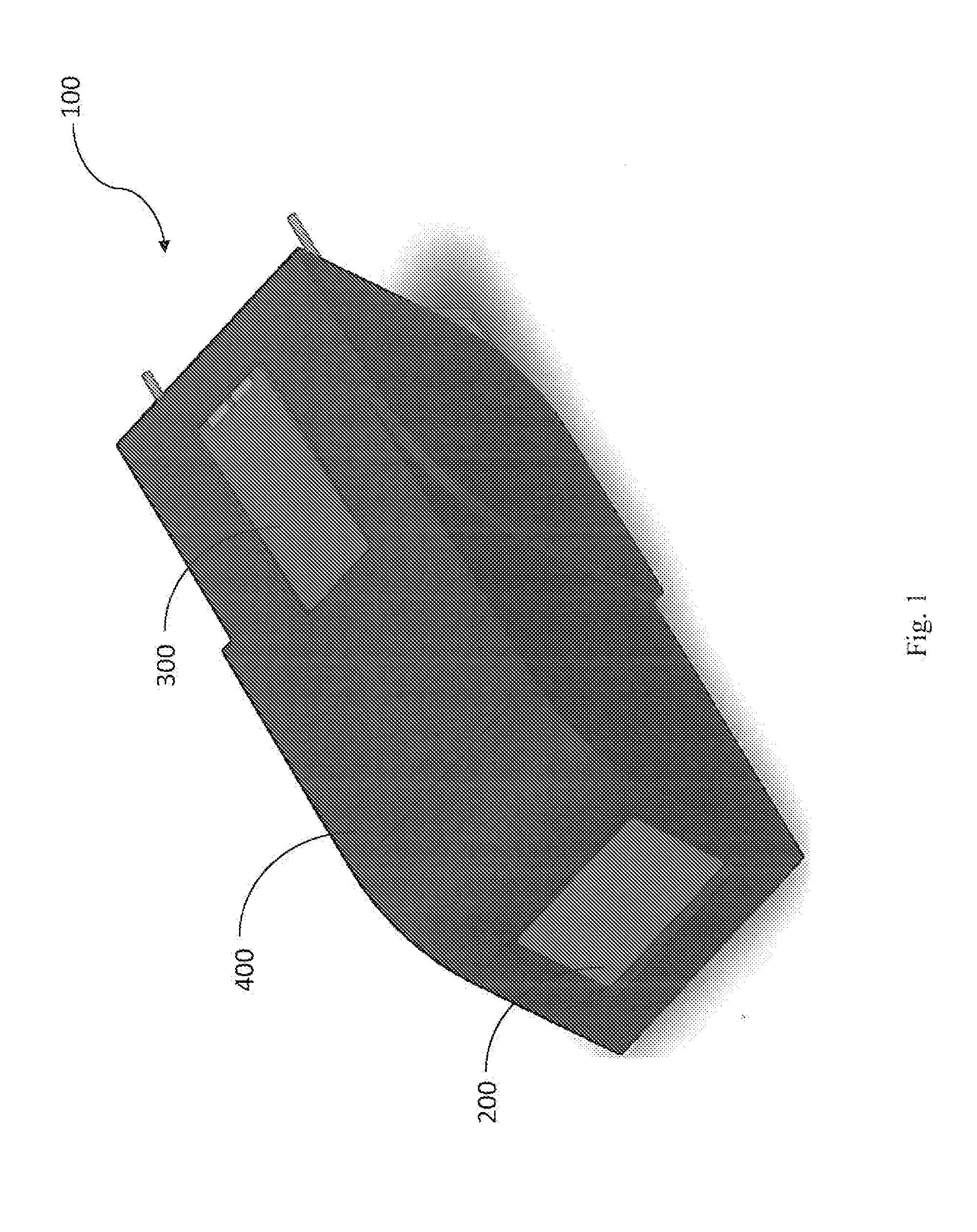
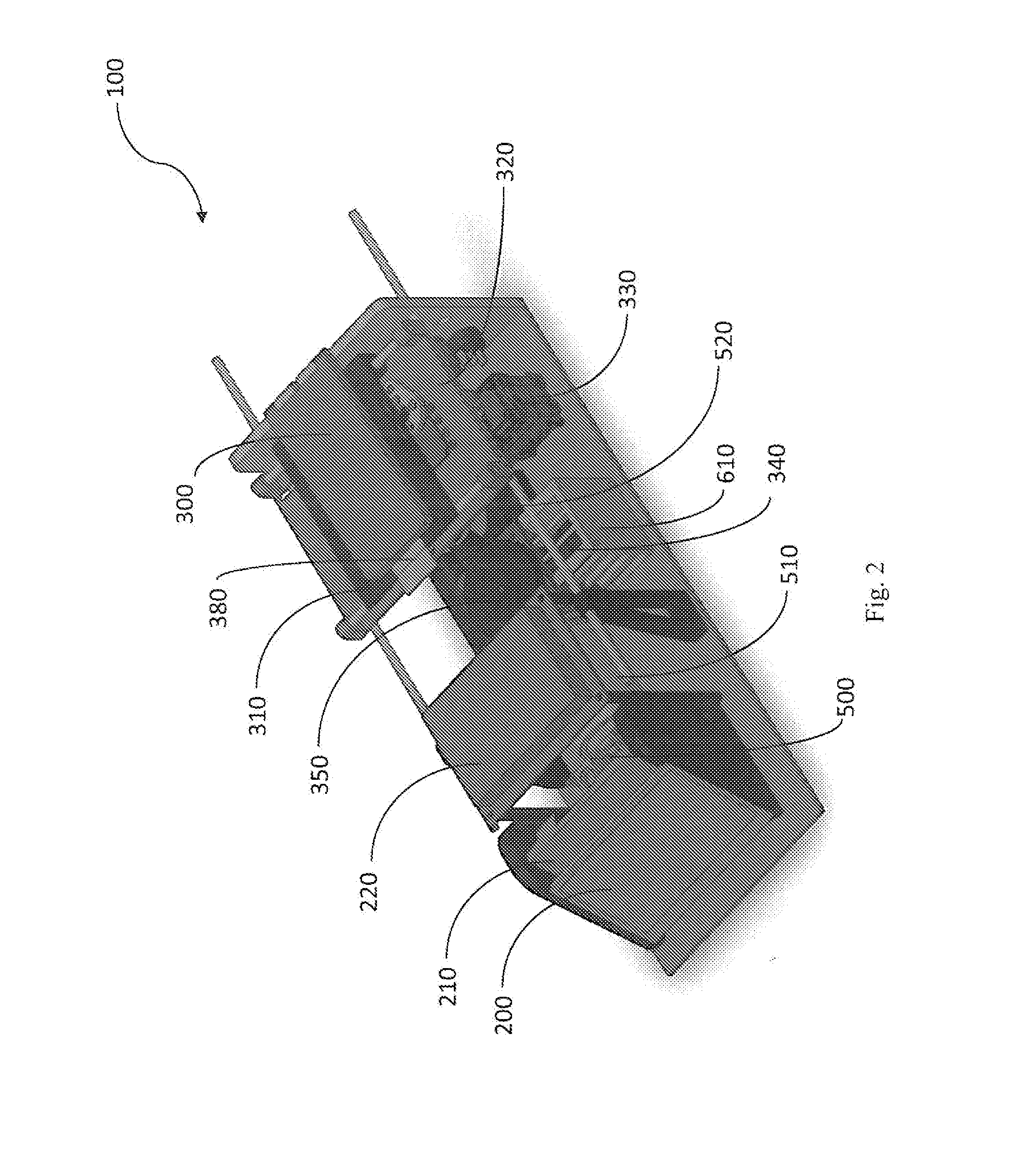
Apparatus and Method for Analyzing a Bodily Sample
a bodily sample and apparatus technology, applied in the field of medical devices, can solve the problems of low throughput, inconvenient process, laborious process, etc., and achieve the effect of reducing the cost of multiple tests and the time consumed for diagnosis, facilitating and expediting the diagnostic procedure, and maintaining the effect of
- Summary
- Abstract
- Description
- Claims
- Application Information
AI Technical Summary
Benefits of technology
Problems solved by technology
Method used
Image

Examples
example 1
[0269]Detection of Trypanosoma Cruzi or Trypanosoma brucei
[0270]Trypanosoma Cruzi is the parasite responsible for the potentially fatal Chaggas disease. One of its life cycle stages (Trypomastigotes) occurs in the blood, where it has a worm-like shape—an elongated body and a flagellum—which constantly twirls and spins in the blood. This motion is the cue for the detection algorithm.
[0271]Human African trypanosomiasis, also termed “African Sleeping Sickness”, is caused by trypanosome parasites Trypanosoma brucei. There is NO immunological or genetic laboratory test for identification of the parasites to date. The software of the invention successfully identified the trypanosome parasites by tracking motion of pixels within successively captured images, in a peripheral blood sample, thus identifying typical motion associated with trypanosome parasites.
[0272]Referring to FIG. 7, an image captured shows Trypanosoma brucei parasites (indicated by arrows), surrounded by red blood cells.
[...
example 2
Detection of Plasmodium and Babesia Species
[0295]Plasmodium are parasites responsible for Malaria disease; Babesia are parasites responsible for Babesiosis disease. Both types of parasites infect red blood cells (RBCs) and in the initial stages of the infection form ring-like structures. Importantly, normal RBCs expel their organelles, and in particular their nucleus, before entering the blood stream. Hence the goal is to detect RBCs which contain a significant amount of DNA, indicating parasitic infection of Plasmodium or Babesia.
[0296]Plasmodium and Babesia species of interest include P. Falciparum, P. Vivax, P. Ovale, P. Malariae, B. Microti and B. Divergens.
Detection Algorithm:
[0297]1. Initially, the blood is stained with fluorochromatic dyes such as Acridine Orange, Giemsa or Ethidium Bromide. Dyes which stain DNA and not RNA or which create a contrast between the two, are preferable.[0298]2. A thin blood smear is prepared upon a cartridge, images are captured, and the images...
PUM
| Property | Measurement | Unit |
|---|---|---|
| depth | aaaaa | aaaaa |
| length | aaaaa | aaaaa |
| diameter | aaaaa | aaaaa |
Abstract
Description
Claims
Application Information