
Method for establishing prediction model based on multidimensional texture of brain nuclear magnetic resonance images

a multi-dimensional texture and brain nuclear magnetic resonance technology, applied in the field of medicine, can solve the problems of difficult for mri technology to evaluate correctly how serious the symptom of ad is, and the only use of hippocampus atrophy as one explanation of the doctor's mri image, and achieve good effects

Active Publication Date: 2016-09-29
CAPITAL UNIVERSITY OF MEDICAL SCIENCES
View PDF13 Cites 9 Cited by
  • Summary
  • Abstract
  • Description
  • Claims
  • Application Information

AI Technical Summary

Benefits of technology

The present invention provides a method for segmenting brain MRI images using region growing and extracting texture features using the Contourlet transform. This method is better than extracting texture by overall segmenting as it preserves margin information. The specificity for predicting sensitivity of early AD is 100% by extracting texture feature parameters from ROIs of brain MRI images. This support vector machine prediction model takes a good effect in early AD added diagnosis.

Problems solved by technology

However, the MRI technology can only use the hippocampus atrophy as one of the indicators to distinguish a patient from a healthy person.
The explanations of doctors to the MRI images are easily affected by subjectivity and lack of consistency.
Moreover, it is hard for the MRI technology to evaluate correctly how serious the symptom of the AD is.
Therefore, there still are many theoretical issues to be studied.
The semi-supervised learning is a key problem studied in the field of pattern recognition and machine learning in recent years, which mainly considers the problem of how to train and classify with a few tag samples and a plurality of samples without tag.

Method used


Image

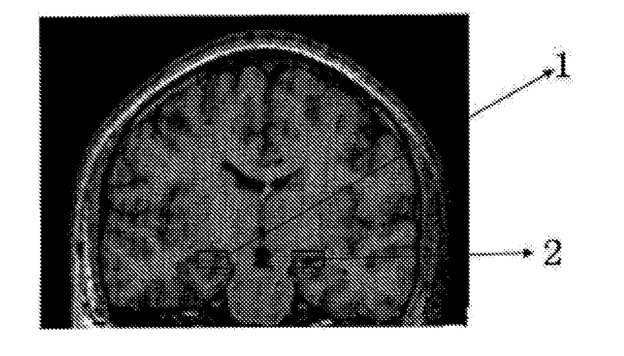
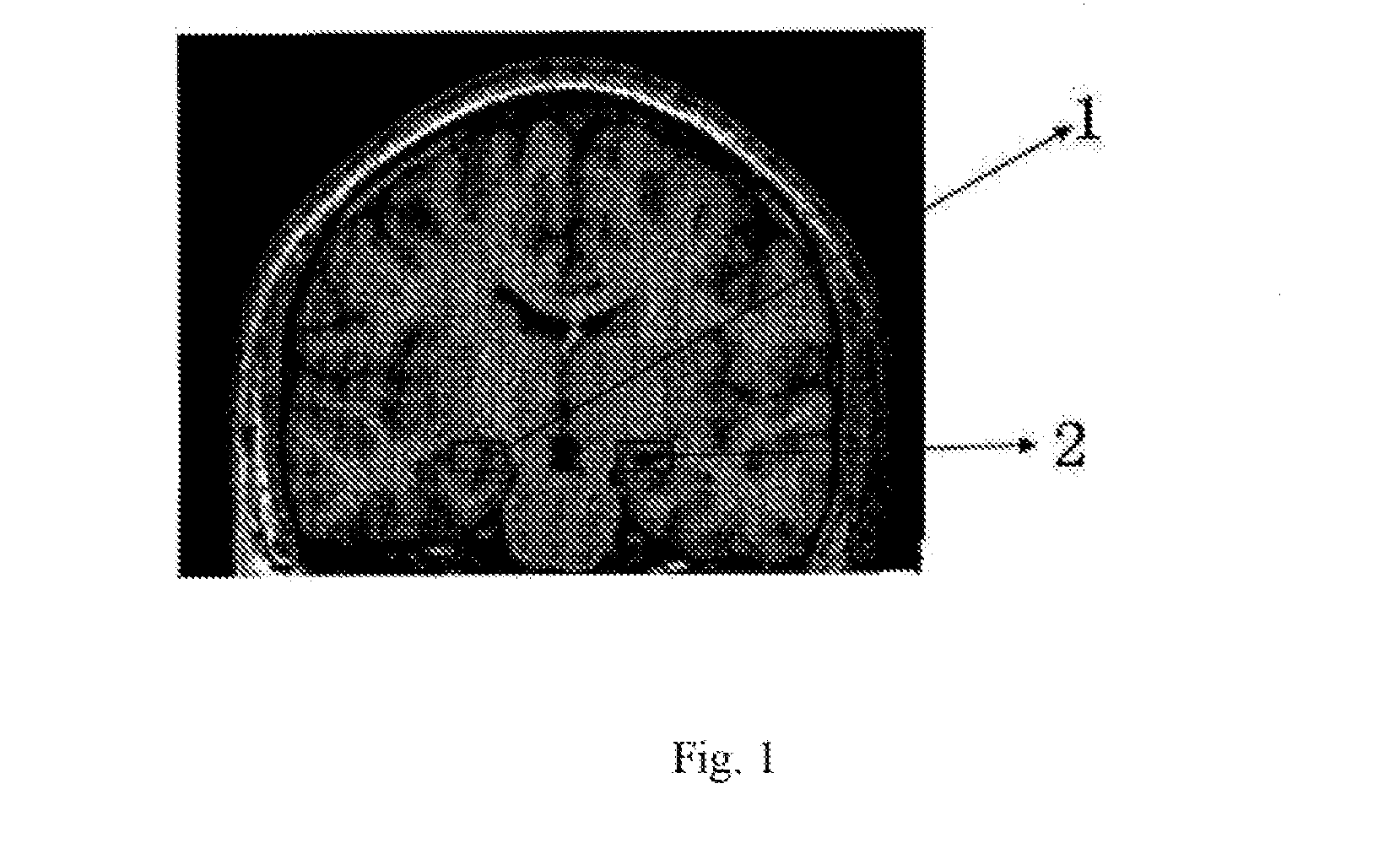
  • Method for establishing prediction model based on multidimensional texture of brain nuclear magnetic resonance images
  • Method for establishing prediction model based on multidimensional texture of brain nuclear magnetic resonance images
  • Method for establishing prediction model based on multidimensional texture of brain nuclear magnetic resonance images

Examples


Embodiment Construction

[0086]The following embodiments are used to describe the model established by using the method of the present invention for predicting AD based on brain MRI images including related ROIs. They are only used to further explain the method of the present invention. However, the embodiments are not used to limit the application scope of the present invention. Actually, the method can also be used to determine properties of other type medical images.

[0087]The source of the images: brain MRI images of the elderly of AD, MCI and the healthy elderly shared on ADNI website, which are in form of .Nii and are read by MRIcro software.

[0088]Methods: programming with Matlab software, segmenting the ROIs in above MRI images with region growing method, and extracting texture feature parameters of related ROIs with Contourlet transform.

[0089]An embodiment for extracting texture feature parameters ROIs of brain MRI images is provided below; its steps are as follows:

[0090]1, respectively collecting a ...


PUM


Abstract

Disclosed is a method for establishing a prediction model based on a multidimensional texture of brain nuclear magnetic resonance images. Images are segmented using a region growing method, a contourlet transform method is used to extract an edge texture feature parameter of ROIs, a multidimensional database is established, and a prediction model is established using various data mining methods, comprising a Gaussian process, a support vector machine, a random forest, a Lasso regression and a semi-supervised support vector machine. The ROIs comprise a hippocampus region and an entorhinal cortex region.

Description

CROSS-REFERENCE TO RELATED APPLICATIONS[0001]This application is a continuation of International Patent Application No. PCT / CN2014 / 001129 with an international filing date of Dec. 16, 2014 designating the United States, now pending and further claims priority benefits to Chinese Patent Application No. 201410023582.5 filed Jan. 17, 2014. The content of the aforementioned applications, including any intervening amendments thereto, is incorporated herein by reference.FIELD OF THE INVENTION[0002]The present application relates to the field of medicine, specifically to a method for establishing a prediction model based on multidimensional textures of brain nuclear magnetic resonance images.BACKGROUND OF THE INVENTION[0003]In aided diagnosis of early Alzheimer disease (AD), it is important to identify features of the ROIs (reactive oxygen intermediates including entorhinal cortex and hippocampus) in the magnetic resonance imaging (MRI) images. However, the MRI technology can only use the ...

Claims


Application Information

Patent Timeline
Patent Type & Authority Applications(United States)
IPC IPC(8): G06T7/00G06T7/40
CPCG06T7/0016G06T7/0081G06T2207/30016G06T2207/10088G06T2207/20141G06T7/403G06T7/0012G06T2207/20016G06T2207/20081G06T7/11G06T7/41G06T7/187
Inventor GUO, XIUHUAGAO, NIWANG, JINGJINGLUO, YANXIAGUO, JIN
Owner CAPITAL UNIVERSITY OF MEDICAL SCIENCES
Learn More