Immunohistochemical proximity assay for pd-1 positive cells and pd-ligand positive cells in tumor tissue

a technology of immunohistochemical proximity and tumor tissue, which is applied in the field of cancer treatment, can solve the problems of significant patient failure to exhibit anti-tumor response, and achieve the effect of facilitating detection and quantification

Active Publication Date: 2016-10-20
MERCK SHARP & DOHME LLC
View PDF1 Cites 34 Cited by
  • Summary
  • Abstract
  • Description
  • Claims
  • Application Information

AI Technical Summary

Benefits of technology

[0005]The inventors have discovered that quantifying the number of PD-1-expressing cells (PD-1+ cells) that are spatially proximal with PD-L1-expressing cells (PD-L1+ cells) in tumor samples is a useful tool to predict which patients are most likely to respond to treatment with a PD-1 antagonist. To facilitate the detection and quantitation of such spatially proximal cells in a tumor sample, the inventors developed a mutliparametric IHC proximity assay, which employs a PD-1-binding antibody, a PD-L1-binding antibody and a random sampling algorithm to generate a proximity score for a tumor tissue section that represents the number of tumor associated pairs of spatially proximal PD-1+ cells and PD-L1+ cells, i.e., cells that are situated within a physiologically relevant distance of each other.

Problems solved by technology

While clinical studies with these antibodies have produced durable anti-tumor responses in some cancer types, a significant number of patients failed to exhibit an anti-tumor response.

Method used


Image

  • Immunohistochemical proximity assay for pd-1 positive cells and pd-ligand positive cells in tumor tissue
  • Immunohistochemical proximity assay for pd-1 positive cells and pd-ligand positive cells in tumor tissue
  • Immunohistochemical proximity assay for pd-1 positive cells and pd-ligand positive cells in tumor tissue

Examples


example 1

Multiplex IHC Assay for PD-1 and PD-L1 Expression in FFPE Tumor Tissue Sections

[0263]This example describes the process used to stain tumor samples for PD-1 and PD-L1 expression that were then imaged and assayed for proximity as discussed in the Examples below.

Reagents

[0264]

WorkingDescriptionCompanyCatalog #Conc.Mouse anti-human PD-L1 mAbIn houseN / A2 ug / mL(Clone 22C3)Envision Flex Mini Kit, High pHDakoK8023N / AEnvision FLEX+ MouseDakoK8022Ready to(LINKER)Use(RTU)Normal Donkey SerumJackson017-000-3 mg / mL1215% byvolumeGoat anti-human PD-1 polyclonalR&DAF10862 ug / mLAbDonkey anti-goat (biotinylated)Jackson705-065-6.5 ug / mL  IgG147ABC EliteVectorPK7100RTUTSA Kit #4 withInvitrogenT-20914See TSAAlexa Fluor 568 tyramideprepTSA Kit #2 withInvitrogenT-20912See TSAAlexa Fluor 488 tyramideprepAntibody DiluentDakoS0809RTUMayer's HematoxylinPolyS216-1GLRTUScientificMicromountLeica3801731RTU

Preparation of Working Reagents from TSA Kit

[0265]Tyramide Stock Solution:

[0266]Dissolved component A into 15...

example 2

Multicolor Immunohistochemistry of Tumor (Melanoma) Samples Demonstrates Distinct Spatial Patterns of PD-1 and PD-L1 Distribution in Archival FFPE Melanoma Samples

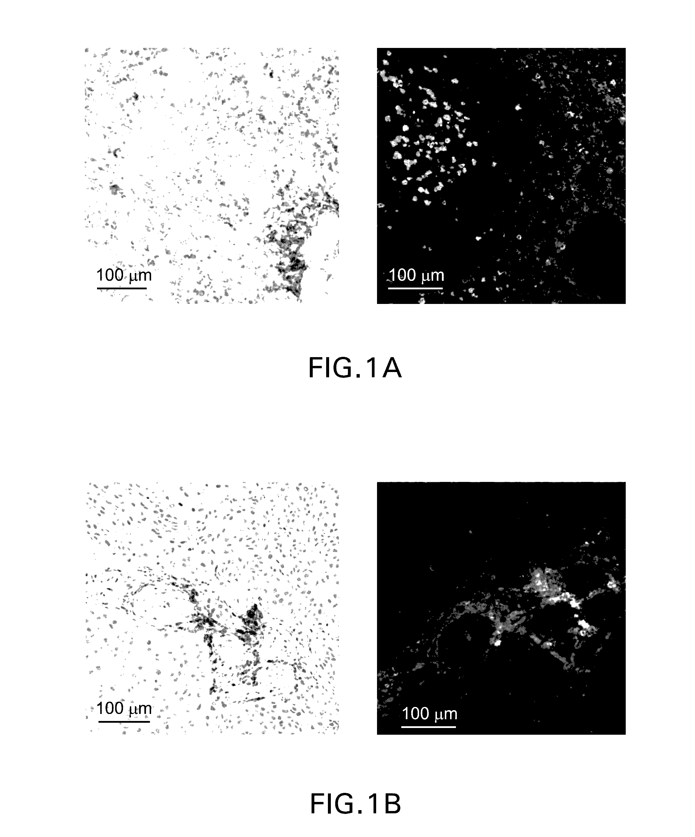
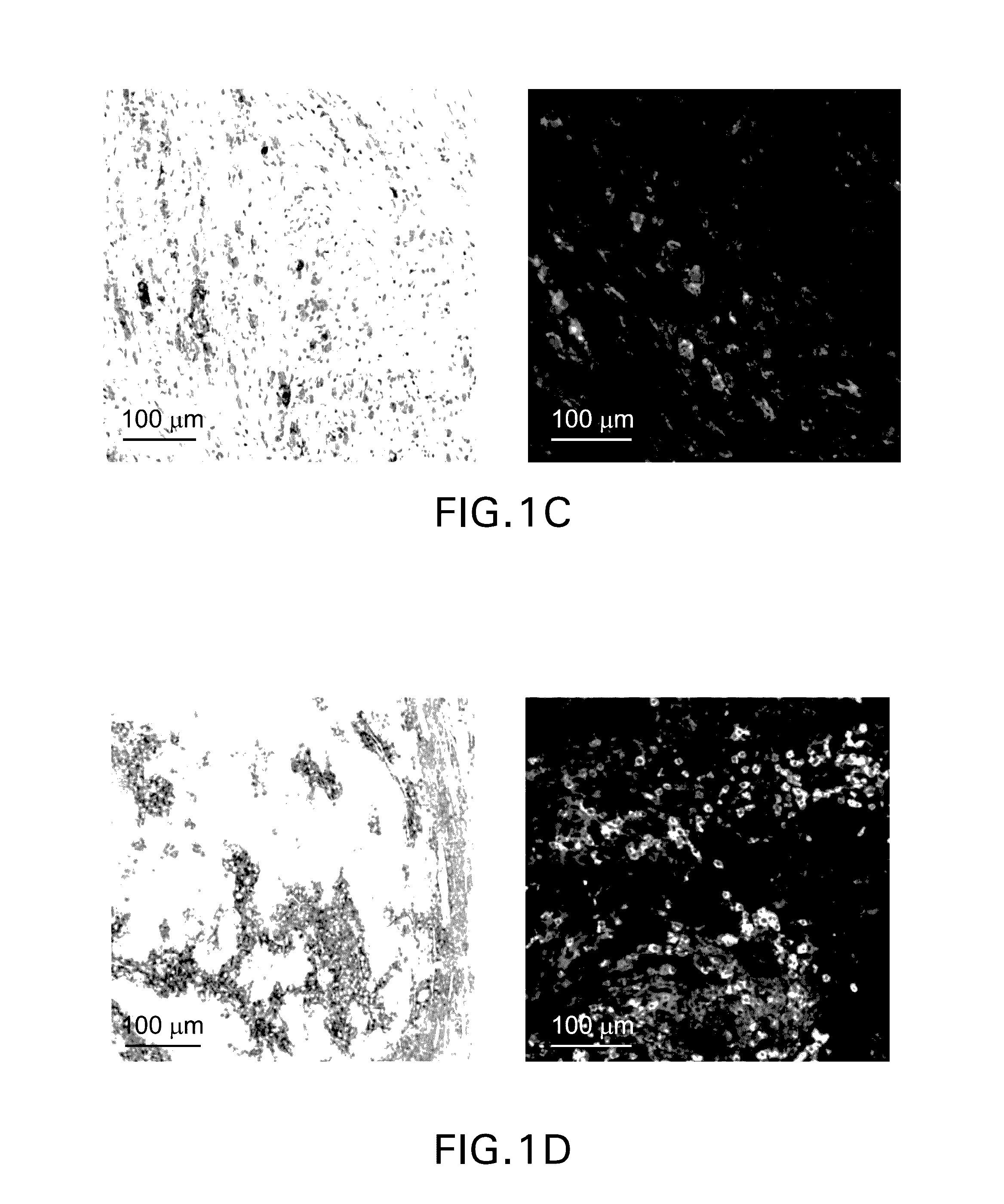
[0276]Single parameter chromogenic immunohistochemical staining was performed using a mouse monoclonal anti-PD-L1 antibody (clone 22C3) and the brown DAB chromogen (FIG. 1, left column) Multiparametric fluorescence immunohistochemistry was performed using the protocol detailed in Example 1. In brief, formalin-fixed paraffin-embedded sections were stained using an anti-PD1 antibody (polyclonal goat anti-hPD-1, green) and an anti-PD-L1 antibody (clone 22C3, red). Representative patterns of PD-1 and PD-L1 are depicted in the images in the right-hand column. In contrast to the single parameter chromogenic stain, which is limited to demonstrating the range of PD-L1 expression per se, the dual fluorescence staining reveals distinct patterns of PD-L1 expression in relation to PD-1+ cells. PD-L1 can be identified as being either (...

example 3

Random Disc Sampling of Tissue Sections for Quantifying PD-1 Expressing Cells and PD-L1 Expressing Cells in Spatial Proximity

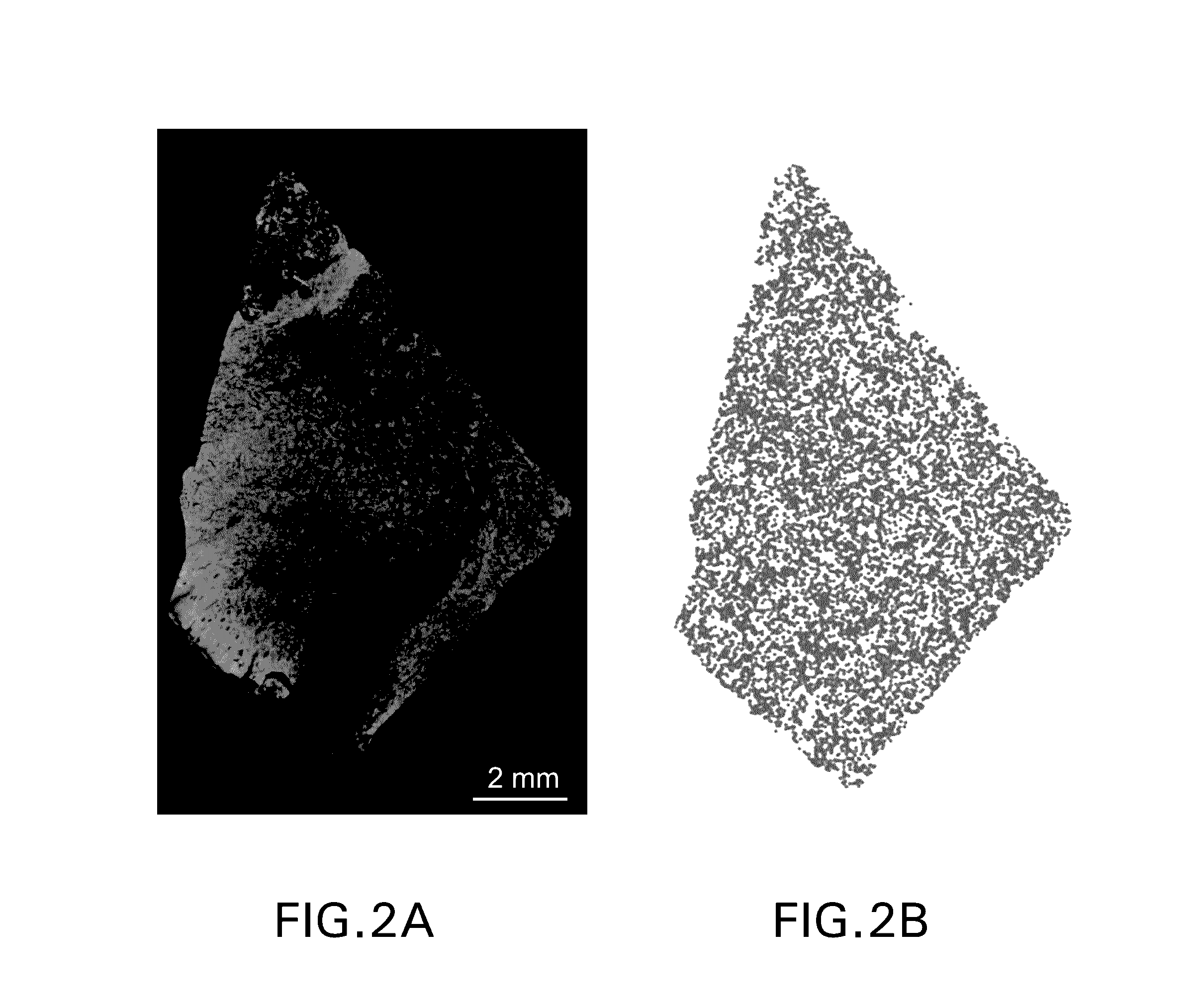
[0277]Multiparametric immunofluorescent immunohistochemistry was performed using the protocol detailed in Example 1. In brief, formalin-fixed paraffin-embedded sections were stained using an anti-PD1 antibody (green) and an anti-PD-L1 antibody (red). The slides were scanned using an Aperio ScanScope FL scanner (20× objective) and captured as afi fused sys files. The images were analyzed for staining artifacts such as the three fluorescent artifacts shown in FIG. 6. If found these artifacts were removed by the intensity distribution of individual channels that correspond to red pixels and green pixels and removed high intensity artifactual fluorescence around the edges of the stained tissue, low intensity artifactual fluorescence across the stained tissue, and used both shape and intensity information to remove the blood artifact. Once these artifacts were remo...


PUM

PropertyMeasurementUnit
diameteraaaaaaaaaa
pHaaaaaaaaaa
threshold proximity scoreaaaaaaaaaa

Abstract

The present disclosure describes an IHC assay for detecting and quantifying spatially proximal pairs of PD-1-expressing cells (PD-1+ cells) and PD-Ligand-expressing cells (PD-L+ cells) in tumor tissue, and the use of the assay to generate proximity biomarkers that are predictive of which cancer patients are most likely to benefit from treatment with a PD-1 antagonist. The disclosure also provides methods for testing tumor samples for the proximity biomarkers, as well as methods for treating subjects with a PD-1 antagonist based on the test results.

Description

FIELD OF THE INVENTION[0001]The present invention relates generally to the treatment of cancer. In particular, the invention relates to the identification of biomarkers for identifying patients whose tumors are likely to respond to treatment with an antagonist of Programmed Death 1 (PD-1).BACKGROUND OF THE INVENTION[0002]PD-1 is recognized as an important player in immune regulation and the maintenance of peripheral tolerance. PD-1 is moderately expressed on naive T, B and NKT cells and up-regulated by TB cell receptor signaling on lymphocytes, monocytes and myeloid cells (1). Two known ligands for PD-1, PD-L1 (B7-H1) and PD-L2 (B7-DC), are expressed in human cancers arising in various tissues, and PD-L1 expression has been associated with poor prognosis and reduced overall survival, irrespective of subsequent treatment, in studies on different cancers, e.g., ovarian, renal, colorectal, pancreatic, liver cancers and melanoma (2-12). Similarly, PD-1 expression on tumor infiltrating l...

Claims


Application Information

Patent Timeline
Patent Type & Authority Applications(United States)
IPC IPC(8): G01N33/574G06T7/40G06T7/00C07K16/28G16B20/00G16B25/10G16H30/40G16H70/60
CPCG01N33/57492G06T2207/20021G06T7/408G06T7/0081G06T7/0012G01N2333/70521G01N2333/70532C07K2317/76A61K2039/505G06T2207/30204G06T2207/30096G06T2207/10056G06T2207/30024G06T2207/10024G06T2207/10064C07K16/2818C12Q1/6886G01N33/574C12Q2600/158G06T7/11G06T7/41G06T7/90G01N2800/52G16H20/40G16H20/17A61P35/00G16H30/40G16H70/60G16B20/00G16B25/10G16B40/20
Inventor PIERCE, ROBERT H.YEARLEY, JENNIFER H.TURNER, SCOTT P.DOGDAS, BELMABAGCHI, ANSUMAN
Owner MERCK SHARP & DOHME LLC
Why Eureka
Try Eureka