Method of imaging multiple retinal structures

a retinal structure and imaging technology, applied in the field of imaging multiple retinal structures, can solve problems such as limiting clinical applications of these methods, and achieve the effect of uniform retinal irradian

Active Publication Date: 2016-11-03
UNIVERSITY OF ROCHESTER
View PDF2 Cites 4 Cited by
  • Summary
  • Abstract
  • Description
  • Claims
  • Application Information

AI Technical Summary

Benefits of technology

[0014]Light may be directed with a light source including a shutter, and the method further comprising controlling the shutter so that light is directed on the portion of the retina only during acquisition of images thereof. The light source may include an acousto-optic modulator, and the method further comprising modulating the power of light directed on the portion of the retina, thereby achieving uniform retinal irradiance.

Problems solved by technology

However, the prior approaches have involved relatively high light levels, focusing challenges and / or relatively long post-processing of image data, thus limiting clinical applications of these methods.

Method used


Image

  • Method of imaging multiple retinal structures
  • Method of imaging multiple retinal structures
  • Method of imaging multiple retinal structures

Examples


Embodiment Construction

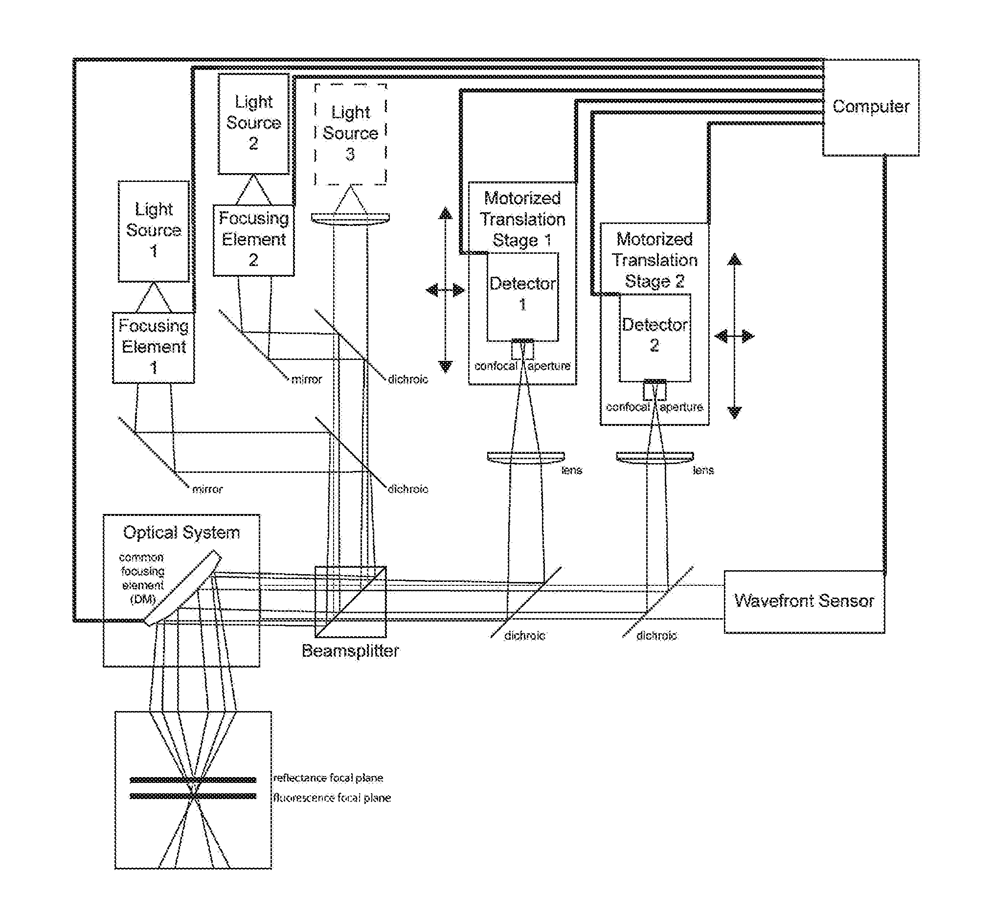
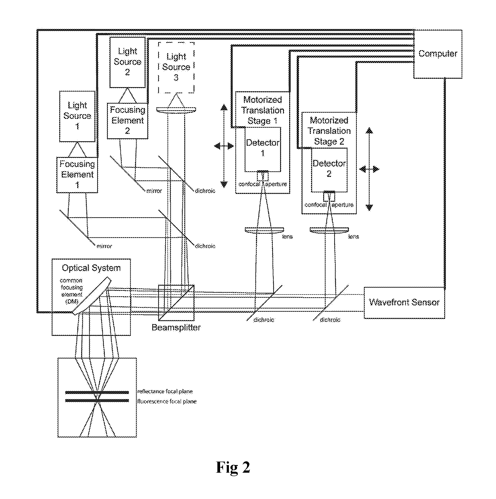
[0044]As discussed above, it has been shown that RPE cells can be imaged with fluorescence AOSLO in normal and diseased eyes. However, high light levels, focusing challenges, and / or long post-processing of data times have limited clinical application of these methods. In particular, the low signal of fluorescence images and the long post-processing time has previously prevented images from being immediately inspected during imaging to ensure that focus, etc., is set appropriately. This invention addresses these problems that hinder routine imaging of the retinal structures in a clinical setting.

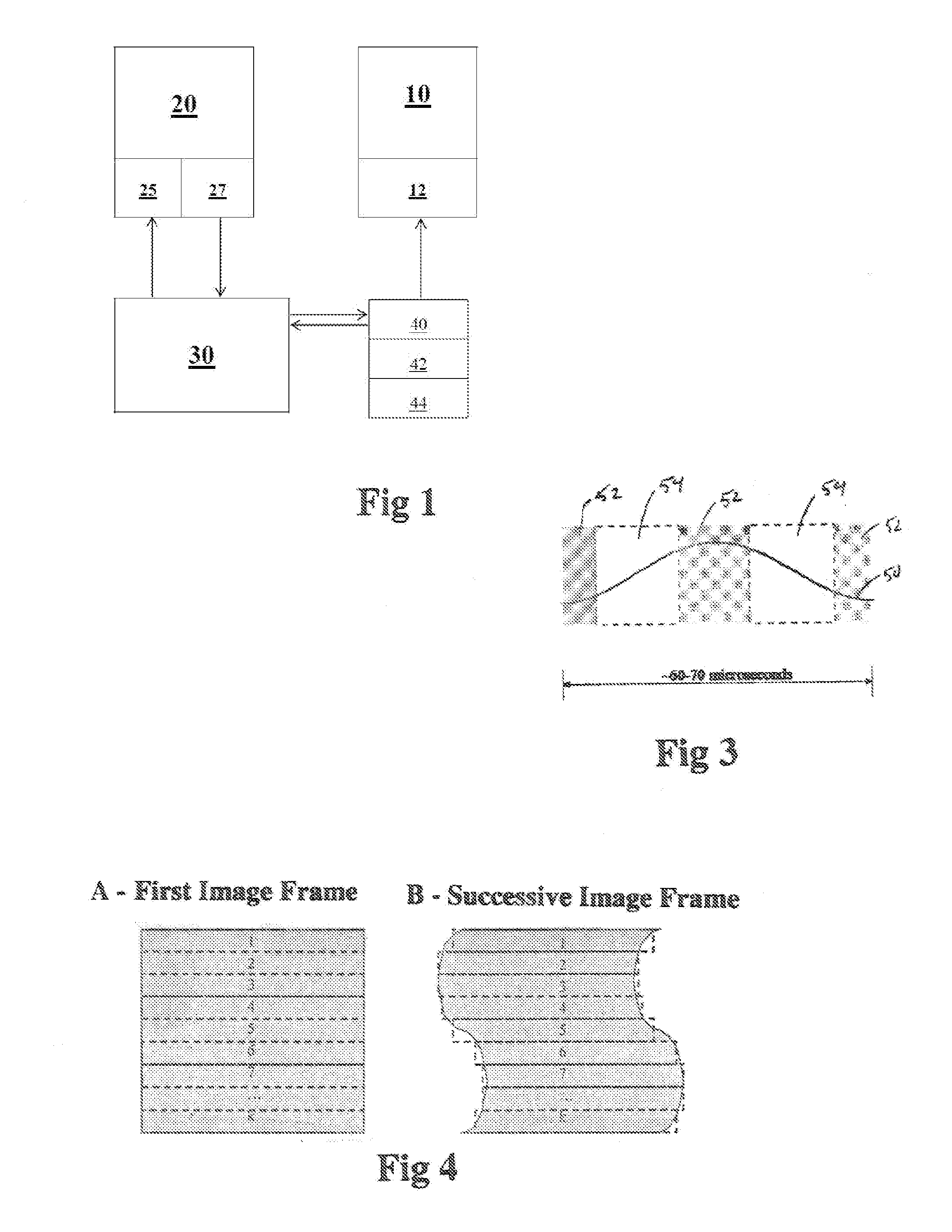
[0045]According to some aspects of this invention, the efficiency of image acquisition and processing is improved, thereby improving the quality of the final composite images while achieving real-time viewing of such final images. A first cross-correlation of images compensates for intra-frame distortion of image frames due to retinal movement during image acquisition. A second cross-correlat...


PUM

PropertyMeasurementUnit
frequencyaaaaaaaaaa
irradianceaaaaaaaaaa
poweraaaaaaaaaa

Abstract

Methods and apparatus for imaging multiple retinal structures and montaging of multiple retinal images are provided. The method involves cross-correlating images from different imaging channels to compensate for intra-frame distortion due to retinal movement during image acquisition, and conducting a second cross-correlation to filter out any motion artifacts in the images. The resultant images are combined to generate a composite image. The method also involves controlling light directed on the retina spatially and temporally.

Description

STATEMENT REGARDING FEDERALLY SPONSORED RESEARCH OR DEVELOPMENT[0001]This invention was made with Government support under Grant Nos. EY014375, and EY001319, awarded by the National Institutes of Health / National Eye Institute. The government has certain rights in the invention.REFERENCE TO A “SEQUENCE LISTING”[0002]Not applicable.BACKGROUND OF THE INVENTION[0003]Non-invasive imaging of various retinal structures, such as retinal pigment epithelial (RPE) cells, of the living eye is important, especially for studying, diagnosing and monitoring diseases of the living human eye. Confocal imaging systems, including those using adaptive optics, have been employed to obtain images of various retinal structures. For example, fluorescence adaptive optics scanning light ophthalmoscope (AOSLO) techniques have been used to image RPE cells in normal and diseased eyes. However, the prior approaches have involved relatively high light levels, focusing challenges and / or relatively long post-process...

Claims


Application Information

Patent Timeline
Patent Type & Authority Applications(United States)
IPC IPC(8): A61B3/12A61B3/00A61B3/10A61B3/13G02B27/00A61B3/14G02F1/11
CPCA61B3/12A61B3/14A61B3/0025G02F1/11A61B3/1015G02B27/0068G02B27/0031A61B3/0058A61B3/1025A61B3/13A61B3/0008G06T2207/30041
Inventor YANG, QIANGROSSI, ETHAN ANDREW
Owner UNIVERSITY OF ROCHESTER
Why Eureka
Try Eureka