Radiographic image processing device, method, and recording medium

a radiation image and processing device technology, applied in the field of radiation image processing devices, can solve the problems of difficult to determine a non-linear gain for preventing overenhancement, difficult to change the pixel value using a linear operation, and difficult to determine a unique gain, etc., to achieve accurate enhancement of the band image, accurate calculation and accurate removal of the scattered radiation componen

Active Publication Date: 2017-03-02
FUJIFILM CORP
View PDF28 Cites 13 Cited by
  • Summary
  • Abstract
  • Description
  • Claims
  • Application Information

AI Technical Summary

Benefits of technology

[0016]Therefore, a method may be considered which calculates a scattered radiation component using a radiographic image that has a linear relationship with the amount of radiation and performs a scattered radiation removal process and a contrast enhancement process. In this case, the scattered radiation component is subtracted from a low-frequency component of the radiographic image and the gain is added to a high-frequency component. In this way, it is possible to enhance contrast while removing scattered radiation.
[0018]The invention has been made in view of the above-mentioned problems and an object of the invention is to provide a technique which can effectively prevent a reduction in contrast caused by scattered radiation of a radiographic image with high accuracy.
[0031]Here, since the second radiographic image has a linear relationship with the amount of radiation, it is possible to accurately calculate the scattered radiation component. As a result, it is possible to accurately remove the scattered radiation component from the second radiographic image. Since the conversion function is calculated from the scattered radiation component of the second radiographic image which has a linear relationship with the amount of radiation, it is possible to accurately enhance the band image using the conversion function. Therefore, in the reconstructed radiographic image, scattered radiation is removed and a high-frequency component included in the radiographic image is enhanced. As a result, a reduction in contrast caused by scattered radiation is prevented.
[0032]In particular, the first radiographic image has a linear relationship with the logarithmic amount of radiation and the pixel value of the band image has a linear relationship with the logarithmic amount of radiation. Therefore, it is possible to easily determine the conversion function from the scattered radiation component. In addition, since the band image is converted by the conversion function, it is possible to effectively enhance a frequency component of the band image. Therefore, it is possible to accurately and effectively prevent a reduction in contrast caused by scattered radiation of a radiographic image.

Problems solved by technology

However, in the radiographic image that has a linear relationship with the amount of radiation, it is difficult to change the pixel value using a linear operation.
Therefore, it is difficult to uniquely determine the gain.
In particular, it is difficult to determine a non-linear gain for preventing over-enhancement in the radiographic image that has a linear relationship with the amount of radiation.
For this reason, when the radiographic image that has a linear relationship with the amount of radiation is used, it is difficult to accurately enhance contrast.
In this case, as described above, since the scattered radiation is a portion of radiation, it is difficult to calculate the scattered radiation component using the radiographic image that has a linear relationship with the logarithmic amount of radiation.

Method used


Image

  • Radiographic image processing device, method, and recording medium
  • Radiographic image processing device, method, and recording medium
  • Radiographic image processing device, method, and recording medium

Examples


first embodiment

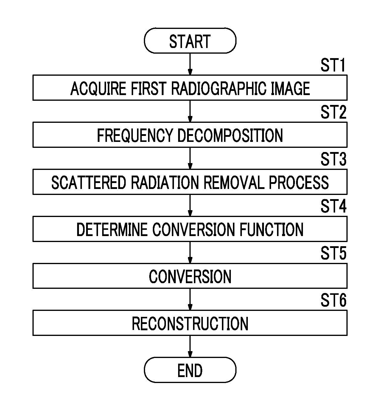
[0087]In the first embodiment, a scattered radiation component is calculated from the radiographic image GL, which is a Gaussian component in the lowest frequency band, and a conversion function map is created. However, a filtering process using a low-pass filter may be performed for the radiographic image G0 to gradate the radiographic image G0, inverse logarithmic conversion may be performed for the gradated radiographic image G0 to acquire a radiographic image GL0 in which a pixel value has a linear relationship with the amount of radiation, and a primary radiation component and a scattered radiation component may be calculated from the radiographic image GL0. In this case, the created conversion function map has the same size as the original radiographic image G0, that is, the Laplacian component L0. Therefore, the conversion function determination unit 24 may reduce the conversion function map (which is referred to as R0 similarly to the above) having the same size as the Lapla...

second embodiment

[0095]In the second embodiment, a storage unit 27 stores the virtual model K of the subject M having the initial body thickness distribution T0 (x, y). The body thickness means the total thickness of a subject region except for an air region on the path of the emitted radiation.

[0096]Next, a body thickness estimation process will be described. FIG. 11 is a flowchart illustrating the body thickness estimation process. The virtual model acquisition unit 31 of the body thickness estimation unit 28 acquires the virtual model K of the subject M having the initial body thickness distribution T0 (x, y) from the storage unit 27 (Step ST31). The virtual model K is data which virtually indicates the subject M and in which a body thickness that follows the initial body thickness distribution T0 (x, y) is associated with each position on an x-y plane. In addition, structures (here, anatomic structures such as a lung field, a bone, and an organ) included in the virtual model K, the arrangement o...

third embodiment

[0118]As such, in the third embodiment, the scattered radiation removal process is performed using the virtual grid characteristics. Therefore, it is possible to obtain the same scattered radiation removal effect as that when imaging is performed using the grid that is assumed to be used.

[0119]In the third embodiment, the scattered radiation transmittance Ts and the primary radiation transmittance Tp are acquired as the virtual grid characteristics. However, only one of the scattered radiation transmittance Ts and the primary radiation transmittance Tp may be acquired.

[0120]In the third embodiment, the radiographic image G0 which is obtained by imaging using a grid may be a processing object. In this case, a process of removing a stripe pattern caused by the grid is performed for the radiographic image G0 and then the scattered radiation removal process is performed for the radiographic image G0. The scattered radiation removal process may acquire a radiographic image (first grid us...


PUM


Abstract

A frequency decomposition unit performs frequency decomposition for a first radiographic image to acquire a plurality of band images. A scattered radiation removal unit calculates a scattered radiation component that is caused by a subject and is included in a second radiographic image which has a linear relationship with the amount of radiation, using the second radiographic image, and removes the scattered radiation component from the second radiographic image to acquire a scattered-radiation-removed radiographic image. A conversion function determination unit determines a conversion function for converting at least one of the band images according to the scattered radiation component included in the second radiographic image. A reconstruction unit converts the band image, using the conversion function, and generates a converted band image. The reconstruction unit reconstructs the scattered-radiation-removed radiographic image and the converted band image to acquire a processed radiographic image.

Description

CROSS REFERENCE TO RELATED APPLICATIONS[0001]The present application is a Continuation of PCT International Application No. PCT / JP2015 / 001211 filed on Mar. 6, 2015, which claims priority under 35 U.S.C. §119(a) to Japanese Patent Application No. 2014-059841 filed on Mar. 24, 2014. Each of the above applications is hereby expressly incorporated by reference, in its entirety, into the present application.BACKGROUND OF THE INVENTION[0002]1. Field of the Invention[0003]The present invention relates to a radiographic image processing device and a radiographic image processing method which perform image processing including a scattered radiation removal process for a radiographic image and a program which causes a computer to perform the radiographic image processing method.[0004]2. Description of the Related Art[0005]In the related art, in the capture of a radiographic image of a subject using radiation that is transmitted through the subject, particularly, when the thickness of the subj...

Claims


Application Information

Patent Timeline
Patent Type & Authority Applications(United States)
IPC IPC(8): A61B6/00G06T7/00
CPCA61B6/5282A61B6/5211G06T2207/10124A61B6/461G06T7/0012A61B6/5235A61B6/483G06T5/002G06T5/50G06T11/005G06T2207/10116G06T2207/20016G06T2207/30004G16H50/20
Inventor KAWAMURA, TAKAHIRO
Owner FUJIFILM CORP
Why Eureka
Try Eureka