
Device and method for a nanofiber wrap to minimize inflamation and scarring

a nanofiber and scarring technology, applied in the field of medical devices, can solve the problems of affecting the regeneration of axonal tissues, and unable to achieve full functional recovery, so as to prevent the infiltration of inflammatory cells and promote healing.

Active Publication Date: 2017-04-06
THE JOHN HOPKINS UNIV SCHOOL OF MEDICINE
View PDF1 Cites 8 Cited by
  • Summary
  • Abstract
  • Description
  • Claims
  • Application Information

AI Technical Summary

Benefits of technology

The present invention is a device made of nanofibers that can promote healing at the connection site between tubular biologic structures. The device consists of a sheet made from a biocompatible material that wraps around the connection site. The sheet is designed to have small pores to prevent inflammatory cells from entering the connection site. This device helps to improve the healing process and make the connection more robust.

Problems solved by technology

Despite great advances in microsurgical techniques, nerve repair continues to be suboptimal, and full functional recovery is seldom achieved.
Research in peripheral nerve injuries has shown that the formation of scar and fibrosis at the site of nerve coaptation impedes axonal regeneration.

Method used


Image

  • Device and method for a nanofiber wrap to minimize inflamation and scarring
  • Device and method for a nanofiber wrap to minimize inflamation and scarring
  • Device and method for a nanofiber wrap to minimize inflamation and scarring

Examples


example

[0028]An exemplary implementation is described herein. This description is merely illustrative and is not meant to be considered limiting. Thy-1 GFP transgenic rats, whose axons constitutively express GFP, were used for the example. Four groups in total were used; 2 for short-term assessment of nerve regeneration measures (Groups 1 and 2) and 2 others for long-term assessment (Groups 3 and 4).

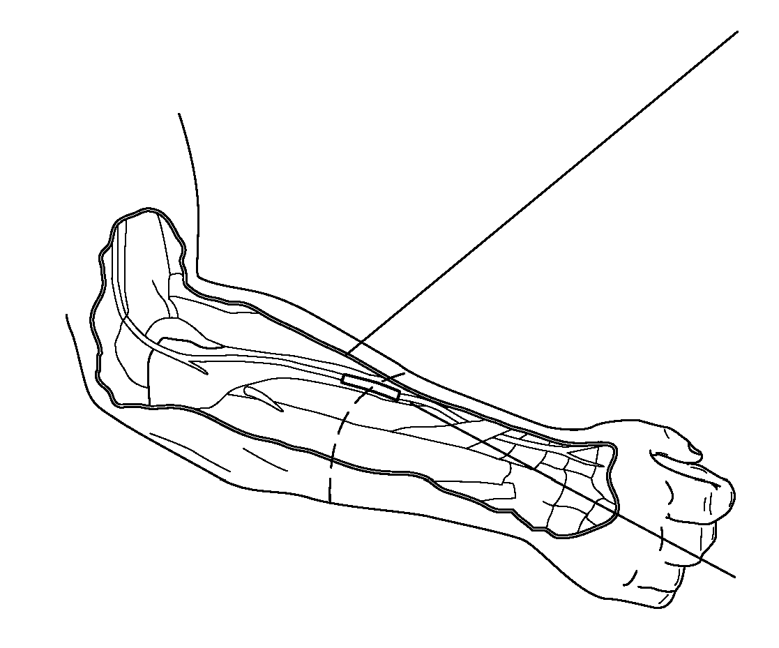
[0029]In Group 1 (control short-term group), a sciatic nerve transection and epineureal repair was performed; in Group 2 (experimental short-term group), in addition to sciatic transection and epineureal repair, the repair site was wrapped with the nanofiber construct. Groups 1 and 2 were harvested at 5 weeks for assessment of nerve inflammation / fibrosis. Groups 3 and 4, consisting of control and experimental long-term groups, respectively, were harvested at 16 weeks for assessment of nerve and muscle functional recovery. FIGS. 4A and 4B illustrate an exemplary rat arm nerve to be wrapped with ...


PUM

PropertyMeasurementUnit
Lengthaaaaaaaaaa
Pore sizeaaaaaaaaaa
Thicknessaaaaaaaaaa

Abstract

The present invention is directed to a device and method for a nanofiber wrap to minimize inflation and scarring of nerve tissue and maximize the nutrient transport. More particularly, the present invention is directed to a novel semi-permeable nanofiber construct prepared from biocompatible materials. The nanofiber construct is applied around a nerve repair site following end-to-end anastomosis. The nanofiber construct is porous and composed of randomly oriented nanofibers prepare using an electrospinning method. The nanofiber construct has a wall that is approximately 50-100 μm thick with pores smaller than 25 μm. The nanofiber construct prevents inflammatory cells from migrating into the nerve coaption site, while still permitting the diffusion of growth factors and essential nutrients. The nanofiber construct allows for enhanced neuroregeneration and optimal function outcomes.

Description

CROSS REFERENCE TO RELATED APPLICATIONS[0001]This application claims the benefit of U.S. Provisional Patent Application No. 61 / 977,881 filed Apr. 10, 2014, which is incorporated by reference herein, in its entirety.FIELD OF THE INVENTION[0002]The present invention relates generally to medical devices. More particularly, the present invention relates to a device and method for a nanofiber wrap to minimize inflammation and scarring of nerve and other tissues as well as any other tubular structure or as a layered constituent of a biologic or synthetic mesh.BACKGROUND OF THE INVENTION[0003]Peripheral nerve injuries constitute a frequent and disabling condition, with an estimated incidence of 23 out of every 100,000 persons per year in developed countries. This statistic does not account for non-traumatic cases (i.e. nerve damage secondary to abdominal or pelvic surgeries) and for lesions not treated at health facilities. Despite great advances in microsurgical techniques, nerve repair c...

Claims


Application Information

Patent Timeline
IPC IPC(8): A61L27/18D01F8/14D01D5/00A61L27/56A61L27/26
CPCA61L27/18A61L27/56A61L27/26D10B2509/00D01F8/14A61L2400/12A61L2430/32D01D5/003A61L27/50C08L67/04C08L89/06
Inventor ZUHAIB, IBRAHIMMAO, HAI-QUANKRICK, KELLINMARTIN, RUSSELLBRANDACHER, GERALD
Owner THE JOHN HOPKINS UNIV SCHOOL OF MEDICINE
Learn More