Method of using an endentulous surgical guide

a surgical guide and endoscope technology, applied in the field of surgical guides, can solve the problems of invasive amount of gum tissue flapping, poor bone quality, and success of dental implants, and achieve the effects of reducing stress on impression copings and connected implants, reducing time, and facilitating the creation of postoperative master models

Active Publication Date: 2017-04-27
IBUR
View PDF0 Cites 36 Cited by
  • Summary
  • Abstract
  • Description
  • Claims
  • Application Information

AI Technical Summary

Benefits of technology

[0016]As the base frame remains in the set position before and after the placement of implants at the surgery, all the superstructures including a transfer attachment will retain the same orientation as they were designed on the preoperative model. Thus, the information obtained by this method is transferable back to the physical or digital preoperative model. Additionally, if this method is used, the surgeon can save time on splinting the implants, and does not have to use an impression material to capture the impression of surrounding gum tissue along with the impression copings since the preoperative model has the all the obtainable information already. This will significantly reduce stress on the impression copings and connected implants.
[0017]The illustrative approaches have significant advantages over conventional teachings. For example, by using a section of the surgical guide set, a postoperative positional index of installed implants will become transferrable and linkable to the digital or physical preoperative model. Since all sections of the surgical guide set are created on the digital preoperative model within the master data file and the design of each s...

Problems solved by technology

Challenges to the successful placement of dental implants include poor bone quality and various hidden anatomical features such as nerves, roots, and sinus cavities.
The primary problem with bone borne surgical guides is the invasiveness of the amount of flapping of the gum tissue that the surgeon has to create in order for the guide to fit correctly on the patient jawbone.
The amount of flapping required increases the likelihood of surgical risks and complications, including blood loss, infection, healing problems, and overall pain experienced by the patient.
Flapping and suturing also requires a great deal of surgical time.
In addition to the problems associated with the surgical procedure, difficulties may also arise when the jawbone has low density, which happens often with Maxilla bones in posterior region.
Low bone density makes it difficult to define the contour of the bone in CT images, which may cause the bone borne surgical drill guide to fit poorly.
Thus, the use of bone borne surgical drill guides has drawbacks and it would be preferred to overcome their associated problems.
While this method is less surgically invasive than the bone borne method, gum tissue borne surgical guides lack stability....

Method used


Image

  • Method of using an endentulous surgical guide
  • Method of using an endentulous surgical guide
  • Method of using an endentulous surgical guide

Examples


Embodiment Construction

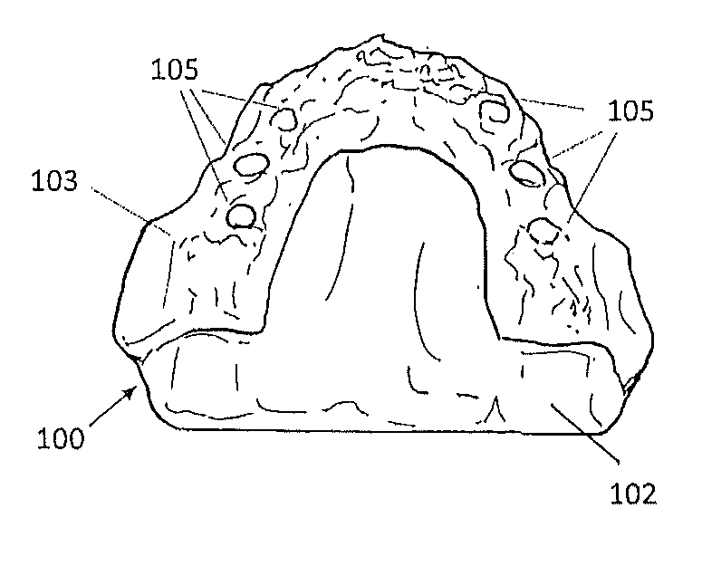
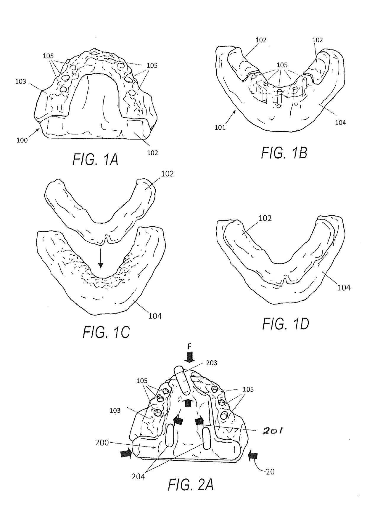
[0087]FIGS. 1A and 1B illustrate patient specific digital or physical dental anatomical diagnostic models 100 and 101 that expose the partial upper jawbone bone structures 103 and a lower jawbone 104 at the surgical sites and the areas of interest. In areas where the bone structures are not exposed, the model has gum tissue surface structures 102. As in FIGS. 1A and 1B, these gum tissue surface structures 102 usually appear towards the distal end of the posterior regions and the palatal area of the upper jaws.

[0088]FIGS. 1C and 1D depict a digital image of a lower jawbone 104 and a digital image of the gum tissue surface structures 102 positioned relative to one another, from which diagnostic anatomical models like 1A and 1B are created.

[0089]The bone structure data can be obtained by tomography imaging devices such as CT and CB CT, and may be exported as a file format, such as STL, suitable for reverse engineering and 3D imaging. Then the data file can be accurately aligned with th...


PUM


Abstract

An approach is disclosed that involves creating an updated master dental model of a patient's mouth after an implant surgery by aligning new postoperative oral scan data to pre-existing preoperative oral scan data that is more comprehensive (e.g., it includes bite registration). Before surgery a multi-piece stackable surgical guide set is created using the preoperative oral scan data. The surgical guide set is used to facilitate the surgery. After the surgery at least one piece of the surgical guide set is placed back in a patient's mouth and may act as a reference marker because it was created using the preoperative oral scan data, but is also part of the postoperative mouth configuration since it was used to facilitate the surgery. The affected portion of the mouth may then be digitally scanned directly or indirectly by way of a physical impression with the reference marker in place to determine the new characteristics resulting from the surgery such as new implant installation locations and orientations. The new postoperative oral scan data may be combined with the pre-existing preoperative oral scan data by way of the reference marker (since it is a constant between the two sets of scan data) without having to do such things as take a new bite registration, on which a new implant restoration can be easily created.

Description

CROSS-REFERENCE TO RELATED APPLICATIONS[0001]This application is a continuation in part application of U.S. patent application Ser. No. 15 / 359,167, filed on Nov. 22, 2016, which is a continuation application of U.S. patent application Ser. No. 13 / 621,146, filed on Sep. 15, 2012, which claims priority to U.S. Provisional Application No. 61 / 535,698, filed on Sep. 16, 2011. This application also claims priority to U.S. Provisional Patent Application No. 62 / 276,083, filed on Jan. 7, 2016. The contents of all of these applications is hereby incorporated by reference in their entirety.FIELD OF THE INVENTION[0002]An improved dental implant method and apparatus is disclosed, and, more particularly, a surgical guide that can be fit to a patient's jawbone.BACKGROUND AND SUMMARY OF THE INVENTION[0003]Dental implants are commonly used in today's dental practices to support various prostheses. Challenges to the successful placement of dental implants include poor bone quality and various hidden ...

Claims


Application Information

Patent Timeline
IPC IPC(8): A61C1/08A61B6/14A61C9/00A61C13/34A61B5/00A61C8/00A61B6/00A61B90/00A61C13/107A61C19/05A61B6/03A61C13/00
CPCA61C1/084A61B6/032A61B6/14A61C9/004A61C13/34A61C13/0004A61B6/4085A61B6/5229A61B90/39A61C13/0001A61C19/05A61B5/4547A61C8/0089B33Y80/00G06T19/20G06T17/00
Inventor GROSCURTH, RANDALL C.GROSCURTH, SHOKO U.
Owner IBUR
Why Eureka
Try Eureka