Systems and methods for detection of structures and/or patterns in images

a system and image technology, applied in the field of image analysis, can solve the problems of limiting the detection technique to automatic nucleus rather than membrane detection, subject to intra- and inter-individual variability, and reducing so as to reduce the computational burden, increase the stability of the cnn, and reduce the number of training images

Active Publication Date: 2017-06-15
VENTANA MEDICAL SYST INC
View PDF4 Cites 116 Cited by
  • Summary
  • Abstract
  • Description
  • Claims
  • Application Information

AI Technical Summary

Benefits of technology

[0019]In accordance with an embodiment of the invention the number of feature maps is below the number of channels of the multi-channel image. This is particularly advantageous for reducing the computational burden and increased stability of the CNN as well as to reduce the number of training images that are required for training the CNN.
[0020]In accordance with a further embodiment of the invention the image sensor that is used to acquire the multi-channel image has a number of color channels that is below the number of channels of the multi-channel image. The co-location data that describes the co-location data that describes the co-location of stains may be utilized for performing the unmixing, such as by using a group sparsity model as it is as such known from the prior art. This way the co-location data can be used both for performing the unmixing and for configuring the CNN.
[0021]The subject disclosure solves the above-identified problems by presenting systems and computer-implemented methods for automatic or semi-automatic detection of structures of interest within images, for example, cellular structures (e.g., cells. nuclei, cell edges, cell membrane), background (e.g., background patterns such as white or white-like space), background image components, and/or artifacts. In exemplary embodiments of the present invention, the present invention distinguishes cellular structures in an image from non-cellular structures or image components. The structures or components may be identified using a convolutional neural network that has been trained for this task. More particularly, the convolutional neural network may be trained to recognize specific cellular structures and features using training images and labels. The neural network outputs a probability that the detected structure does in fact represent a cell, membrane, background, etc. These probabilities may undergo a local maxima finding method such as non-maximum suppression in order to identify a particular pixel that will be used as the “location” of the object. A particular part of the cell, e.g., the approximate center of a nucleus, is illustratively used as the “location” of the object within the area under observation, i.e. an image patch.
[0022]Operations described herein include retrieving individual color channels from a multi-channel image and providing said multiple individual channels as input for a detector, for example, a cell detector. The

Problems solved by technology

This is an extremely tedious and time consuming process and the results may also subject to intra- and inter-individual variability.
However, these techniques are limited to automatic nucleus detection rather than membrane detection.
Although some machine l

Method used


Image

  • Systems and methods for detection of structures and/or patterns in images
  • Systems and methods for detection of structures and/or patterns in images
  • Systems and methods for detection of structures and/or patterns in images

Examples


Embodiment Construction

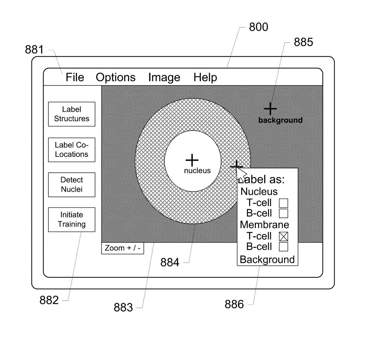
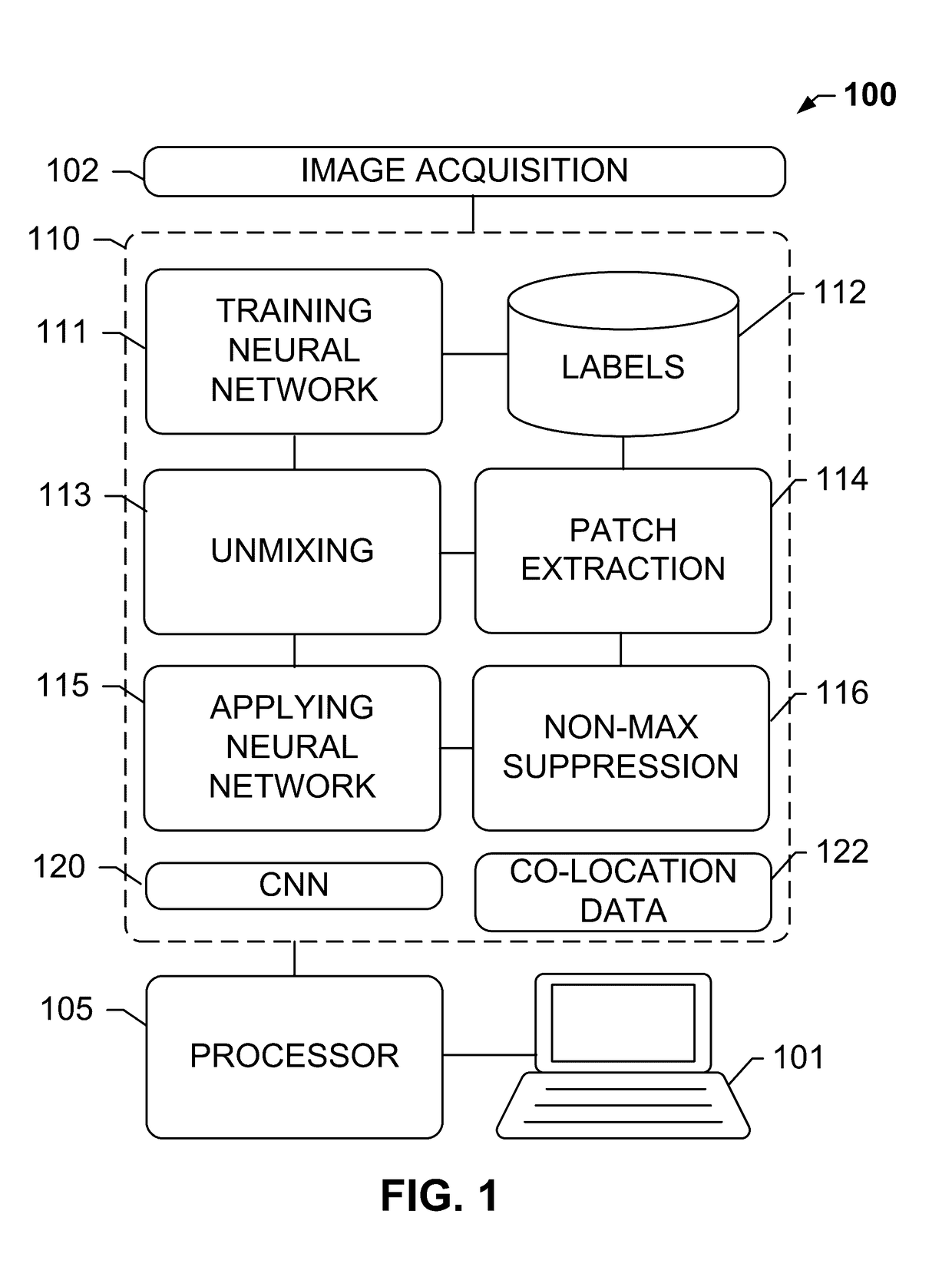
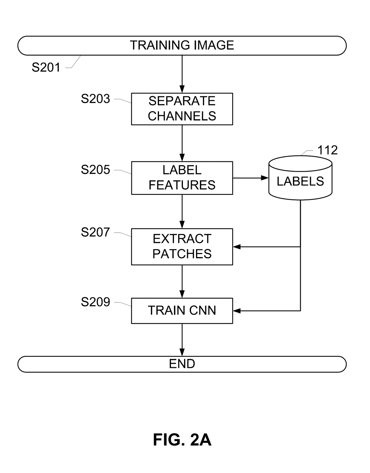
[0036]The subject disclosure solves the above-identified problems by presenting systems and computer-implemented methods for automatic detection of image structures, for example, cellular structures, including retrieving individual color channels from a multi-channel image and providing one or multiple individual channels or portions of image data from the one or more multiple individual channels as input for a cell detector that is trained using a convolutional neural network to identify the immune cells in one or multiple channels of the image that corresponds to an immune cell marker or other target structure such as a nucleus. The multi-channel image may be an RGB image obtained from a brightfield scanner, an image from another color space such as Lab, a multi-channel image from a multi-channel brightfield or darkfield scanner, a fluorescent image from a multi-spectral imaging system, a darkfield image, or any other multi-channel image. In some embodiments the image may be an im...


PUM


Abstract

The subject disclosure presents systems and computer-implemented methods for automatic immune cell detection that is of assistance in clinical immune profile studies. The automatic immune cell detection method involves retrieving a plurality of image channels from a multi-channel image such as an RGB image or biologically meaningful unmixed image. A cell detector is trained to identify the immune cells by a convolutional neural network in one or multiple image channels. Further, the automatic immune cell detection algorithm involves utilizing a non-maximum suppression algorithm to obtain the immune cell coordinates from a probability map of immune cell presence possibility generated from the convolutional neural network classifier.

Description

RELATED APPLICATIONS[0001]This patent application is a continuation of International Patent Application No. PCT / EP2015 / 061226 filed May 21, 2015, which claims priority to and the benefit of U.S. Provisional Application No. 62 / 098,087, filed Dec. 30, 2014, and entitled “SYSTEMS AND METHODS FOR DEEP LEARNING FOR CELL DETECTION”, and to U.S. Provisional Application No. 62 / 002,633, filed May 23, 2014, and entitled “DEEP LEARNING BASED AUTOMATIC CELL COUNTING SYSTEM AND METHOD”, the contents of which are hereby incorporated by reference herein in their entirety into this disclosure.BACKGROUND OF THE SUBJECT DISCLOSURE[0002]Field of the Subject Disclosure[0003]The present subject disclosure relates to image analysis. More particularly, the present subject disclosure relates to automatically identifying structures (e.g., cellular structures) or patterns (e.g., background or white space) in an image.[0004]Background of the Subject Disclosure[0005]In the analysis of biological specimens such...

Claims


Application Information

Patent Timeline
IPC IPC(8): G06T7/00G06K9/00
CPCG06T7/0012G06K9/00127G06T2207/20081G06T2207/30024G06T2207/10024G06T2207/20084G06N3/084G06V20/69G06V20/698G06N3/045
Inventor CHEFD'HOTEL, CHRISTOPHECHEN, TING
Owner VENTANA MEDICAL SYST INC
Why Eureka
Try Eureka