Autoantibody detection method, method for testing possibility of autoimmune disease contraction, autoantibody detection reagent, and autoimmune disease test reagent

a technology for autoantibody detection and autoimmune diseases, applied in the field of autoantibody, can solve the problems of false negative results and inability to detect autoantibody in all patients, and achieve the effects of high specificity, high accuracy and high accuracy

Active Publication Date: 2017-06-22
OSAKA UNIV
View PDF0 Cites 4 Cited by
  • Summary
  • Abstract
  • Description
  • Claims
  • Application Information

AI Technical Summary

Benefits of technology

[0010]It is known that, in an antigen-presenting cell, a protein antigen is degraded into peptide fragments, which are then bound to MHC class II molecules and carried to the surface of the cell, whereby the peptide fragments are presented on the surface of the cell. However, through diligent research, the inventors of the present invention made the following findings. Specifically, the inventors of the present invention first discovered that an MHC class II molecule not only presents the degraded peptide fragment, but also binds to a denatured protein resulting from misfolding in an endoplasmic reticulum (ER) and presents the denatured protein on the cell surface. The inventors of the present invention further discovered that an autoantibody recognizes and binds to the denatured protein presented on the cell surface by the MHC class II molecule. These mechanisms had not been reported at the time the present application was filed, and the inventors of the present invention first discovered these mechanisms. The inventors of the present invention conducted experiments to detect various autoantibodies using, as an antigen reagent, a purified protein used in a conventional ELISA method as described above (i.e., a correctly folded protein resulting from correct folding) or the denatured protein / MHC class II. As a result, the inventors of the present invention verified that the denatured protein / MHC class II is recognized by autoantibodies of autoimmune disease patients with higher specificity as compared with the correctly folded protein, thereby establishing the present invention. Therefore, according to the present invention in which the denatured protein / MHC class II is used as an antigen reagent for detecting an autoantibody, it is possible to detect the autoantibody involved in an autoimmune disease with high accuracy. Also, the present invention can detect autoantibodies undetectable by the ELISA method in which a correctly folded protein is used, for example. Accordingly, the present invention can inhibit the conventional problem of a false negative, thus allowing the possibility of an autoimmune disease to be determined with high accuracy. Therefore, the present invention is very useful in the fields of clinical practice and biochemistry, for example.
[0011]The reason why an autoantibody can be detected with higher accuracy by using the denatured protein / MHC class II as an antigen reagent as compared with the case where the correctly folded protein is used as an antigen reagent presumably is as follows. That is, it is presumed that an autoantibody in the sample binds to a correctly folded protein through cross-reactivity, whereas the autoantibody specifically binds to the denatured protein / MHC class II. It is to be noted, however, that this presumption does not limit the present invention by any means.

Problems solved by technology

However, even when patients are diagnosed with an autoimmune disease by doctors on the basis of their symptoms, the autoantibody is not necessarily detected in all the patients by the ELISA method.
Thus, if diagnosis is made depending on the detection of the autoantibody by the ELISA method alone, false negative results may be obtained.

Method used


Image

  • Autoantibody detection method, method for testing possibility of autoimmune disease contraction, autoantibody detection reagent, and autoimmune disease test reagent
  • Autoantibody detection method, method for testing possibility of autoimmune disease contraction, autoantibody detection reagent, and autoimmune disease test reagent
  • Autoantibody detection method, method for testing possibility of autoimmune disease contraction, autoantibody detection reagent, and autoimmune disease test reagent

Examples


example 1

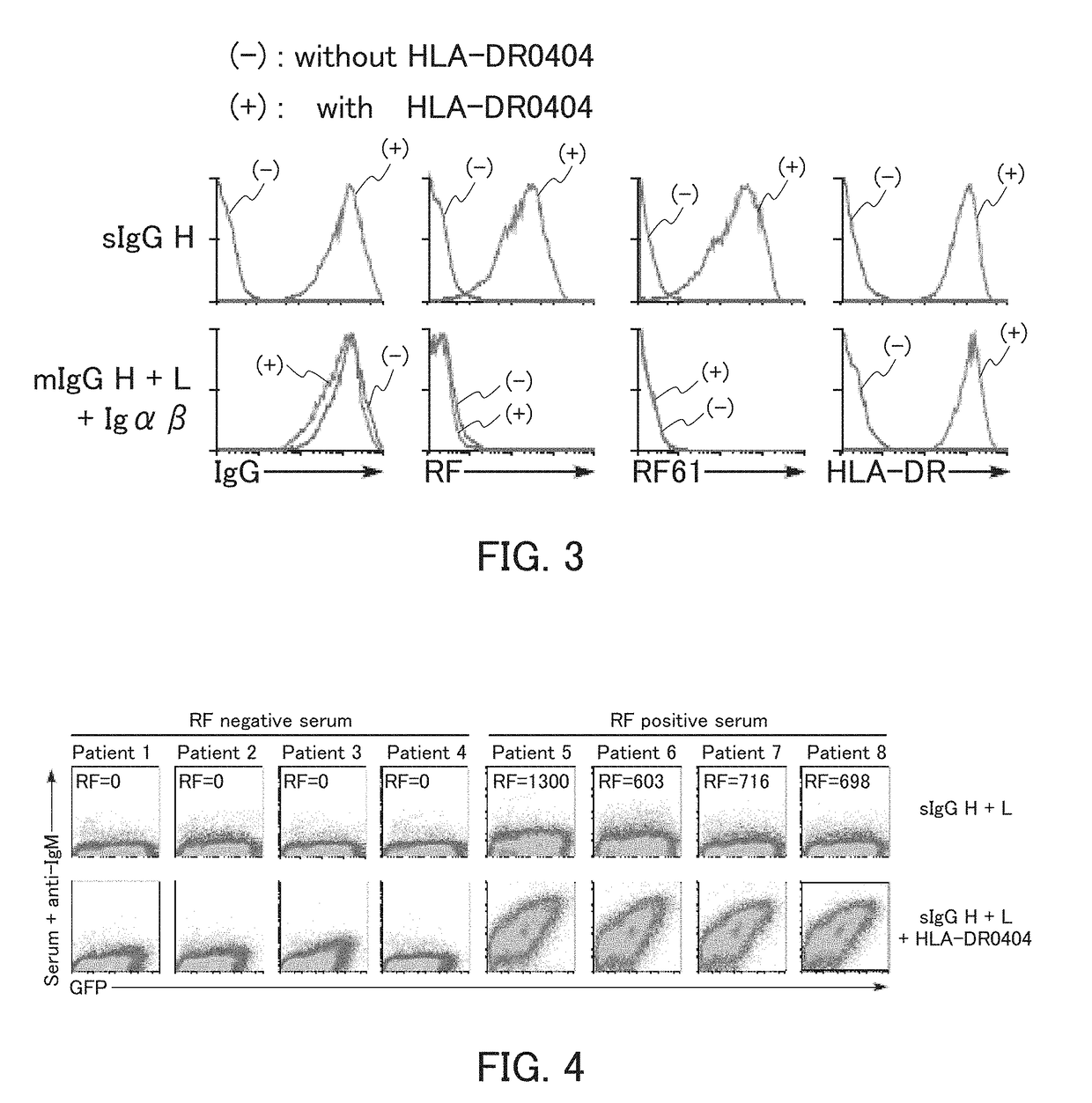
[0136]Example 1 relates to the detection of an autoantibody as an indicator of rheumatoid arthritis.

example 1a

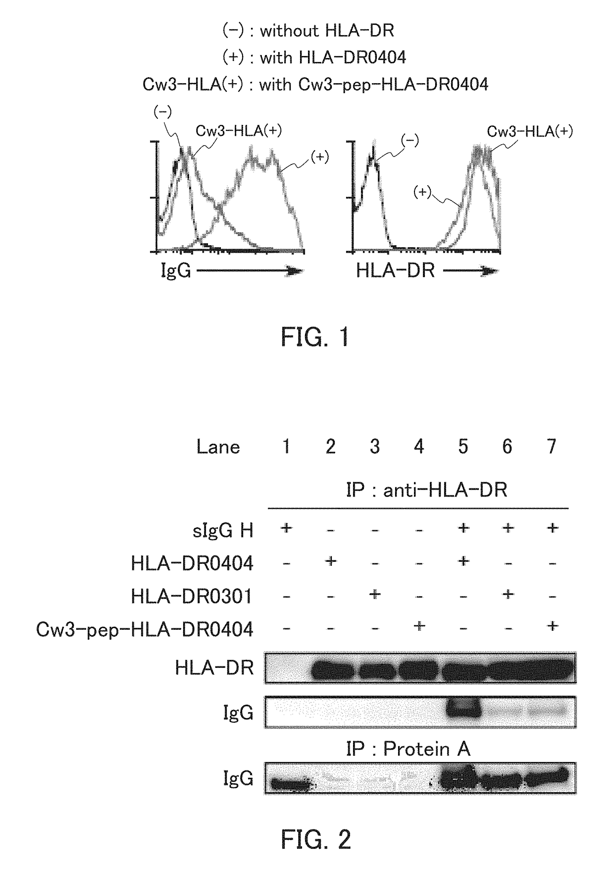
[0137]In the present example, HLA-DR and an IgG heavy chain were expressed, and whether the IgG heavy chain was presented on cell surfaces by the HLA-DR was examined.

[0138]The HLA-DRA*01:01 vector as an α-chain expression vector, the HLA-DRB1*04:04 vector as a β-chain expression vector, the sIgGH vector for secretory IgG heavy chain expression, and the GFP vector were introduced to 293T cells, and the 293T cells were cultured. The cultured cells were reacted with an allophycocyanin (APC)-labeled anti-human IgG Fc antibody or anti-HLA-DR antibody, and further reacted with an APC-labeled anti-mouse IgG antibody. Thereafter, the cells were subjected to flow cytometry analysis. Specifically, using a flow cytometer (trade name; FACS Calibur™, Becton Dickinson), IgG heavy chain expression and HLA-DR expression on surfaces of GFP-positive cells were examined. As Control 1, the analysis was performed in the same manner, except that the HLA-DR expression vectors were not introduced. As Contr...

example 1b

[0143]In the present example, HLA-DR was immunoprecipitated, and whether an IgG heavy chain was bound to the peptide-binding groove of the HLA-DR was examined.

[0144]To 293T cells, the HLA-DRA*01:01 as an α-chain expression vector and the other respective expression vectors were introduced so as to achieve the combinations shown in FIG. 2 to be described below. The 293T cells were then cultured. The cultured cells were lysed in a 0.5% NP-40 solution (polyoxyethylene(9)octyiphenyl ether), and the resultant cell lysate was immunoprecipitated using a biotinylated anti-HLA-DR antibody and streptavidin sepharose (GE Healthcare).[0145]biotinylated anti-HLA-DR antibody for immunoprecipitation: Clone L243, available from ATCC, mAb

[0146]Western blotting was performed on the immunoprecipitated sample. Specifically, the sample was applied to electrophoresis, and IgG and HLA-DR were detected using a peroxidase-labeled anti-human IgG antibody or a rabbit anti-HLA-DR a antibody and a peroxidase-la...


PUM

PropertyMeasurementUnit
temperatureaaaaaaaaaa

Abstract

The present invention provides an autoantibody detection method that can detect an autoantibody causing an autoimmune disease with high accuracy. The autoantibody detection method of the present invention includes the steps of; causing a sample and an antigen reagent comprising a denatured protein presented by an MHC class II molecule to come into contact with each other; and detecting a complex of an autoantibody in the sample and the denatured protein in the antigen reagent. By detecting a complex of the autoantibody and the denatured protein in a biological specimen isolated from a subject according to this detection method, it is possible to test the possibility of the autoimmune disease in the subject from the detection result.

Description

TECHNICAL FIELD[0001]The present invention relates to an autoantibody detection method, a method for testing the possibility of an autoimmune disease, an autoantibody detection reagent, an autoimmune disease test reagent, a method for producing an autoantibody detection reagent, and a screening method for an antigen protein against an autoantibody relevant to an autoimmune disease.BACKGROUND ART[0002]Diagnosis of an autoimmune disease generally is done utilizing, in addition to direct determination based on the symptoms etc. of a patient, indirect determination based on the detection of an autoantibody specific to the autoimmune disease. The detection of an autoantibody usually is performed using, e.g., an ELISA method in which a purified antigen protein is immobilized on a carrier and the binding of the autoantibody with the immobilized protein is examined. However, even when patients are diagnosed with an autoimmune disease by doctors on the basis of their symptoms, the autoantibo...

Claims


Application Information

Patent Timeline
Patent Type & Authority Applications(United States)
IPC IPC(8): G01N33/564G01N33/569
CPCG01N33/56977G01N33/564
Inventor ARASE, HISASHITANIMURA, KENJIJIN, HUIARASE, NORIKO
Owner OSAKA UNIV
Why Eureka
Try Eureka