
Medical device and methods of use

a medical device and ureteroscope technology, applied in the field of medical devices, can solve the problems of loss of kidneys, inability to continue to break stones or stone fragments into smaller fragments without repositioning the ureteroscope, and small stone fragments not passing naturally

Active Publication Date: 2017-08-03
BOSTON SCI SCIMED INC
View PDF10 Cites 9 Cited by
  • Summary
  • Abstract
  • Description
  • Claims
  • Application Information

AI Technical Summary

Benefits of technology

The present invention provides devices and methods for breaking an object into smaller particles and removing them from the human body. The methods involve positioning a distal end of a medical device near the object, applying suction through a lumen of an elongate member to seal the object, and moving the elongate member to remove the sealed object. The devices may include a tube, an elongate member, and a laser fiber or other imaging device. The technical effect is improved devices and methods for breaking and removing objects from the human body.

Problems solved by technology

An impacted kidney stone is a source of intense pain and bleeding, a source of infection and, if a stone completely blocks the flow of urine for any extended length of time, can cause the loss of a kidney.
Intense light energy from a laser within the ureteroscope breaks the stone into increasingly smaller pieces However, in some cases, the stone and / or the stone fragments may be pushed away from the ureteroscope by the laser, thus making it impossible to continue to break the stone or stone fragments into smaller fragments without repositioning the ureteroscope.
However, in some cases, these small stone fragments may not pass naturally.
The combined cross-sectional areas of these two elements may make a device too large to reach the target kidney stone.
Often, the space within the kidney and / or lower calyx of the kidney is more limited than the space within the ureter and this space may not be large enough to accommodate both a working channel for a laser fiber and a lumen for applying suction.

Method used


Image

  • Medical device and methods of use
  • Medical device and methods of use
  • Medical device and methods of use

Examples


Embodiment Construction

[0023]Reference is now made in detail to examples of the present disclosure, examples of which are illustrated in the accompanying drawings. Wherever possible, the same reference numbers will be used throughout the drawings to refer to the same or like parts. The term “distal” refers to a position farther away from a user end of the device. The term “proximal” refers a position closer to the user end of the device. As used herein, the terms “approximately” and “substantially” indicate a range of values within + / −5% of a stated value.

[0024]Overview

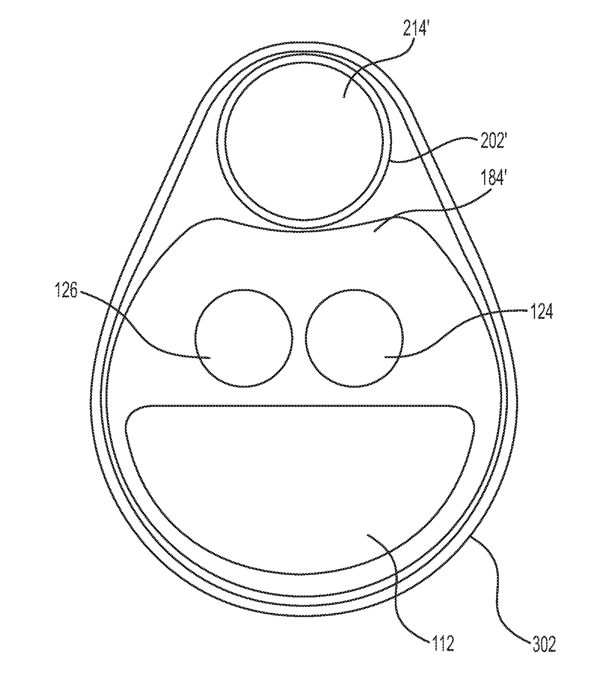
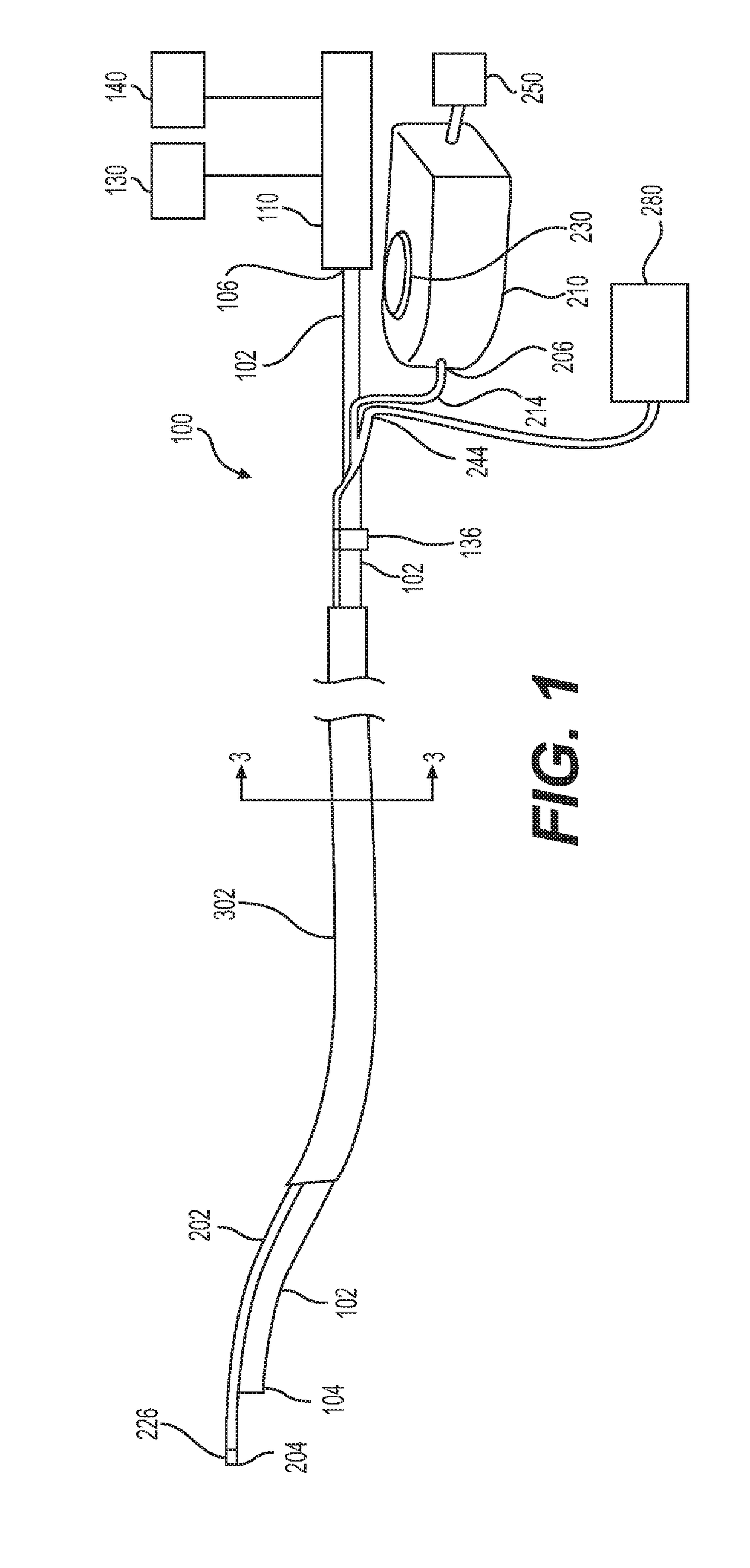
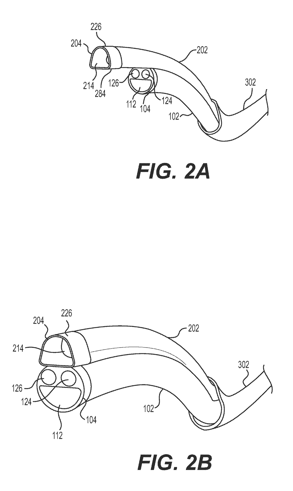
[0025]Aspects of the present disclosure relate to systems and methods for breaking kidney stones into smaller particles and removing those particles from a target area of a patient's body. The medical device described herein may work by securing an elongate member for suction to an outer surface of a tube of a delivery device. A distal end of the tube and a distal end of the elongate member may be positioned within a ureter of a patient. In...


PUM


Abstract

The present disclosure is directed to a medical device. Systems and methods are provided for utilizing a laser to break a kidney stones into smaller fragments and / or dust, and removing particles, stone fragments and / or stone dust from a patient. The medical device may include a delivery device including a tube, and an elongate member having a distal end, a proximal end, and a lumen extending between the proximal end and the distal end, wherein the elongate member is configured to move axially relative to the tube and apply suction through the distal end.

Description

CROSS-REFERENCE TO RELATED APPLICATIONS[0001]This patent application claims the benefit of priority under 35 U.S.C. §119 to U.S. Provisional Patent Application No. 62 / 288,734, filed Jan. 29, 2016, which is herein incorporated by reference in its entirety.TECHNICAL FIELD[0002]The present disclosure relates generally to medical devices. More particularly, the disclosure relates to medical devices used, for example, in breaking objects into smaller particles, and removing the resulting particles from a patient. The disclosure also relates to methods of using such devices.BACKGROUND[0003]The incidence of hospitalization for the removal of urinary calculi, commonly referred to as kidney stones, has been estimated at 200,000 cases per year. A vast majority of these patients pass their stones spontaneously; however, in the remainder, the kidney stone(s) become impacted in the ureter, a muscle tube joining the kidney to the bladder. An impacted kidney stone is a source of intense pain and b...

Claims


Application Information

Patent Timeline
IPC IPC(8): A61B17/22A61B18/24A61B17/30A61B18/26
CPCA61B17/22A61B18/26A61B18/245A61B90/30A61B2017/306A61B2018/00511A61B90/361A61B17/30A61B1/00094A61B1/00135A61B1/015A61B1/307A61B2018/00982A61M2205/106A61M2210/1082A61M1/84A61M1/85
Inventor HARRAH, TIMOTHY P.OSKIN, CHRISTOPHER L.LENZ, DERRICKBANERJEE, ARPITAGAVADE, SANDESHTAKALE, ABHIJITMISRA, PAVAN
Owner BOSTON SCI SCIMED INC
Learn More