Device and method for determining image quality of a radiogram image

Active Publication Date: 2017-08-10
KONINKLJIJKE PHILIPS NV
View PDF0 Cites 13 Cited by
  • Summary
  • Abstract
  • Description
  • Claims
  • Application Information

AI Technical Summary

Benefits of technology

[0008]There may be a need to improve syste

Problems solved by technology

When acquiring chest radiographs, patient positioning errors are a common course of inad

Method used


Image

  • Device and method for determining image quality of a radiogram image
  • Device and method for determining image quality of a radiogram image
  • Device and method for determining image quality of a radiogram image

Examples


Embodiment Construction

[0042]The illustration in the drawings is purely schematic and does not intend to provide scaling relations or size information. In different drawings or figures, similar or identical elements are provided with the same reference numerals. Generally, identical parts, units, entities or steps are provided with the same reference symbols in the description.

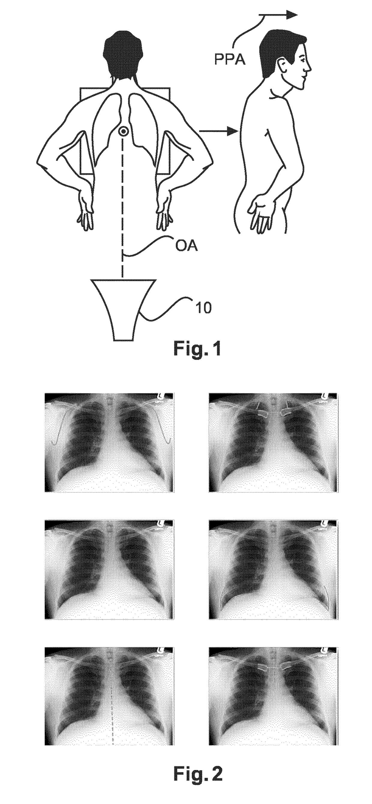
[0043]FIG. 1 shows a position required for an optimal patient's position for a chest PA radiograph, PA: radiation from posterior to anterior. The relative position of a patient to an optical axis OA of an X-ray camera of an acquiring module 10 is shown in FIG. 1.

[0044]In other words, the acquiring module 10 may comprise an X-ray camera or an X-ray detector or any other optical instrument that records images per radiography that can be stored directly.

[0045]The positioning of the patient may be defined by a position vector PPA describing the position of the patient with respect to a reference system or as a relative position with res...


PUM


Abstract

The present invention relates to a device (1) for determining image quality of a radiogram image, the device comprising: an acquiring module (10), which is configured to acquire the radiogram image; a segmentation module (20), which is configured to segment a thoracic bone structure on the acquired radiogram image into segmented thoracic bone structure contours and a tissue structure on the acquired radiogram image into segmented tissue structure contours; and a quality-determination module (30), which is configured to determine a image quality coefficient for the acquired radiogram image based on a comparison of a first position of the segmented thoracic bone structure contours with a second position of the segmented tissue structure contours.

Description

FIELD OF THE INVENTION[0001]The present invention relates to the field of image quality analysis, determination, and assessment. Particularly, the present invention relates to a device and a method for determining image quality of a radiogram image.BACKGROUND OF THE INVENTION[0002]U.S. Pat. No. 6,516,046 B1 describes a method for exact positioning of a patient for radiotherapy or radiosurgery comprising the steps of pre-positioning the patient relative to a linear accelerator, producing at least one X-ray image of the patient or one of body parts in the vicinity of the radiation treatment target, mapping the X-ray image and detecting a positional error on the basis of specific landmarks in the image.[0003]U.S. 2003 / 0081734 A1 describes a low-dose exposure aided positioning system for digital radiography, a system and a method for improved imaging of a patient through the use of low-dose exposure aided positioning is provided, the patient is positioned in the X-ray system and then im...

Claims


Application Information

Patent Timeline
IPC IPC(8): A61B6/00G06T7/62G06T7/00
CPCA61B6/5217G06T7/0014G06T7/62G06T2207/30061A61B6/544G06T2207/10124A61B6/466A61B6/488A61B6/505G06T2207/30008G06T2207/30168G06T7/12G16H50/30
Inventor VON BERG, JENSYOUNG, STEWARTCAROLUS, HEIKE
Owner KONINKLJIJKE PHILIPS NV
Why Eureka
Try Eureka