MRI protocol for segmentation of an image detail using images acquired at two different magnetic field strengths

a magnetic resonance imaging and image detail technology, applied in the field of magnetic resonance imaging protocol for segmentation of image detail, can solve the problem of labour-intensive manual segmentation of the hippocampus from magnetic resonance images, and achieve the effect of accurately segmenting pre-determined image details

Active Publication Date: 2017-08-31
KONINKLJIJKE PHILIPS NV
View PDF1 Cites 5 Cited by
  • Summary
  • Abstract
  • Description
  • Claims
  • Application Information

AI Technical Summary

Benefits of technology

[0004]An object of the invention is to provide a magnetic resonance imaging protocol that more accurately segments pre-determined image details, in particular the hippocampus from surrounding brain structure, from a magnetic resonance image.
[0017]Thus, the image information of an image detail in the training images assists to improve the reliability of the segmentation of a corresponding image detail in the diagnostic image. Training images from several atlases of training image can be used. Thus, in the framework of the present invention, higher main magnetic field strength may represent ultra-high magnetic field (7 T and higher), while lower main magnetic field strength may indicate high magnetic field strength of 3 T, or a commonly used magnetic field strength of 1.5 T. The magnetic resonance imaging protocol of the invention may be formed as a method that has groups of methods steps for acquisition of magnetic resonance signal, groups of method steps for reconstruction of one or more magnetic resonance images from the magnetic resonance signal and groups of method steps for segmentation of a detail, such as substrucure within an an organ of interest, from the magnetic resonance images and further groups of method steps may be included form image processing such as image registration steps. In practice the magnetic resonance imaging protocol may be implemented in software, i.e. in the form of a computer programme that has groups of instructions to run the groups of method steps of the magnetic resonance imaging protocol of the invention. The groups of instructions or method steps that each form a partial set of instructions for the magnetic resonance imaging protocol of the invention form segments, such as acquisition segments, reconstruction segments and segmentation segments that each contain groups of method steps or instructions that pertain to the groups of activities, acquisition, reconstruction and image processing such as segmentation and registration of the magnetic resonance imaging protocol of the invention. These segments each relate to a consistent part of the magnetic resonance imaging protocol each with distinct input and output. The output of one segment may in general from input for a next segment.
[0024]The model-based segmentation makes use of an atlas of the training magnetic resonance images. A global registration of the training magnetic resonance images is made to the diagnostic magnetic resonance image. The training magnetic resonance image is registered to the diagnostic magnetic resonance image. Thus, the highest image quality viz. that of the training image, is maintained for the diagnostic images; registration most often reduces image quality e.g. because of image interpolation artefacts. A volume-of-interest (VOI) is selected around the image detail, e.g. the hippocampus, from the registered diagnostic image and the corresponding volume of interested is also selected from the training magnetic resonance image(s). For example, on each hemisphere of the brain, i.e., there are two volumes-of-interest VOIs (left, right hemispheres) per brain volume. Then the volume-of-interest in the training magnetic resonance images is registered with respect to the corresponding volume-of-interest in the diagnostic magnetic resonance image. The selected volume of interest in the diagnostic image and the corresponding volume of interest in the training image pertain to regions around the corresponding image details, such as e.g. the hippocampus, in the diagnostic magnetic resonance image and in the training magnetic resonance image. On the basis of the registered volume-of-interest, the surface of the image detail (e.g. the surface of the hippocampus) is aligned with the corresponding image detail in the training magnetic resonance images. The segmentation proper is applied e.g. employing an energy minimisation, which is known per se in the field of image segmentation, to the aligned image detail. In fact, the image detail being more clearly represented in the training image assists in aligning the image detail in the diagnostic magnetic resonance image so that the segmentation proper is so well initialised that the image detail is accurately segmented, even if it is less well represented in the diagnostic magnetic resonance image. In this way the segmentation of the image detail from the diagnostic image benefits from the better image quality of the training magnetic resonance image, without the need for higher main magnetic field strength for the acquisition of the diagnostic magnetic resonance image. Notably, the magnetic resonance training magnetic resonance images are acquired at a high magnetic field strength at 7 T and its level of detail facilitates the accurate segmentation of notably the hippocampus from diagnostic magnetic resonance images at lower field strength of 3 T.
[0032]These parameters of the acquisition sequence achieve, when applied at the main magnetic field strength of 3 T, that the reconstructed magnetic resonance image that is reconstructed from the acquired magnetic resonance signal resembles an magnetic resonance image at a higher magnetic field strength of e.g. 7 T of the same anatomy. Thus, the diagnostic magnetic resonance images are acquired such that they resemble the training magnetic resonance images. Thus, in the diagnostic magnetic resonance image (based on data acquired at the lower main magnetic field strength of e.g. 3 T) the way image details are represented is closely to the way such image details are represented in the training images (based on data acquired at the higher main magnetic field strength of e.g. 7 T). This leads to good results in accurate segmentation of the image detail from the diagnostic magnetic resonance image supported by the use of the training magnetic resonance image.
[0033]The echo-train length (ETL) and the parallel imaging (PI) (e.g. SENSE) reduction factor is a compromise between short echo train length which reduces signal loss due to decay of the signal along the echo-train and loss of signal-to-noise ratio, due to the undersampling of k-space at higher parallel imaging reduction facto, at higher parallel imaging (PI) reduction factor. As usual, this reduction factor represents the degree of undersampling in k-space of the magnetic resonance signal relative the sampling density that satisfies the Nyquist criterion to avoid signal aliasing. The short echo train length renders the acquisition sequence less sensitive to motion (e.g. cardiac motion or respiratory motion) in the part of the patient to be examined to be examined. Good results are achieved by a modest undersampling along the first phase encode direction (e.g. reduction factor of 1.4) and a higher reduction for the second phase encode direction (e.g. reduction factor of 2.0). The first and second phase encoding direction are mutually orthogonal in k-space. Furthermore the ETL and echo-time are controlled by half-Fourier. Therefore, the optimal setting according to this aspect of the invention is a compromise of both SENSE and half-Fourier, resulting in the optimum echo-time and ETL. Notably, the choice of the echo time TE and the repetition time TR achieve a sufficient SNR of the acquired magnetic resonance signal, even such that only a single signal average (NSA=1) is sufficient. This limits the total signal acquisition time.

Problems solved by technology

Because manual segmentation of the hippocampus from magnetic resonance images is labour-intensive, automated segmentation is needed for clinical use.

Method used


Image

  • MRI protocol for segmentation of an image detail using images acquired at two different magnetic field strengths
  • MRI protocol for segmentation of an image detail using images acquired at two different magnetic field strengths
  • MRI protocol for segmentation of an image detail using images acquired at two different magnetic field strengths

Examples


Embodiment Construction

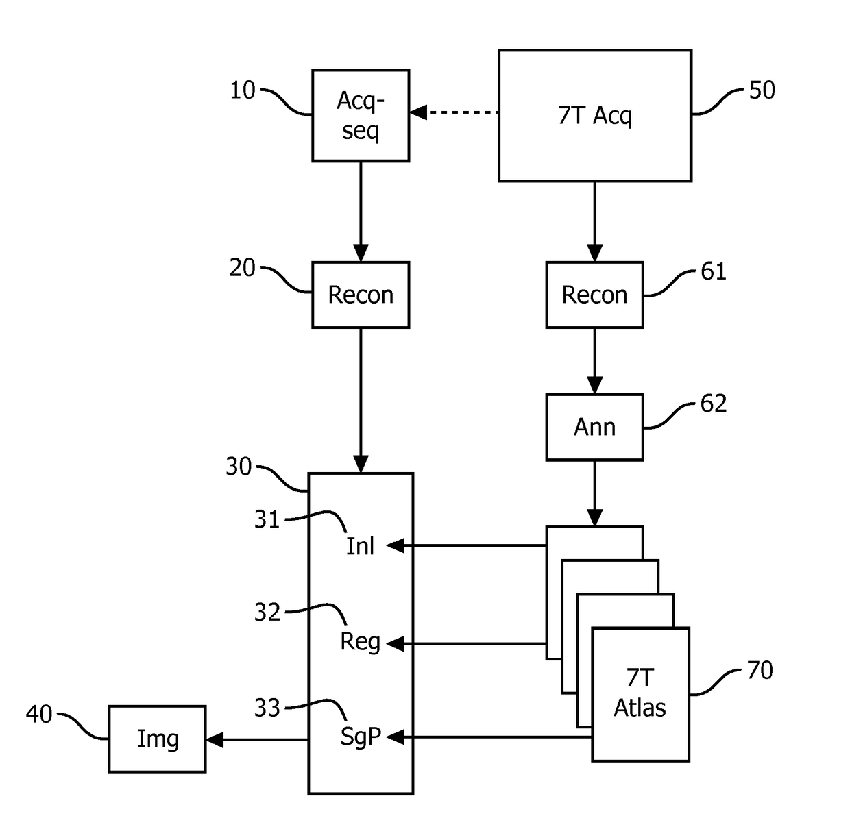
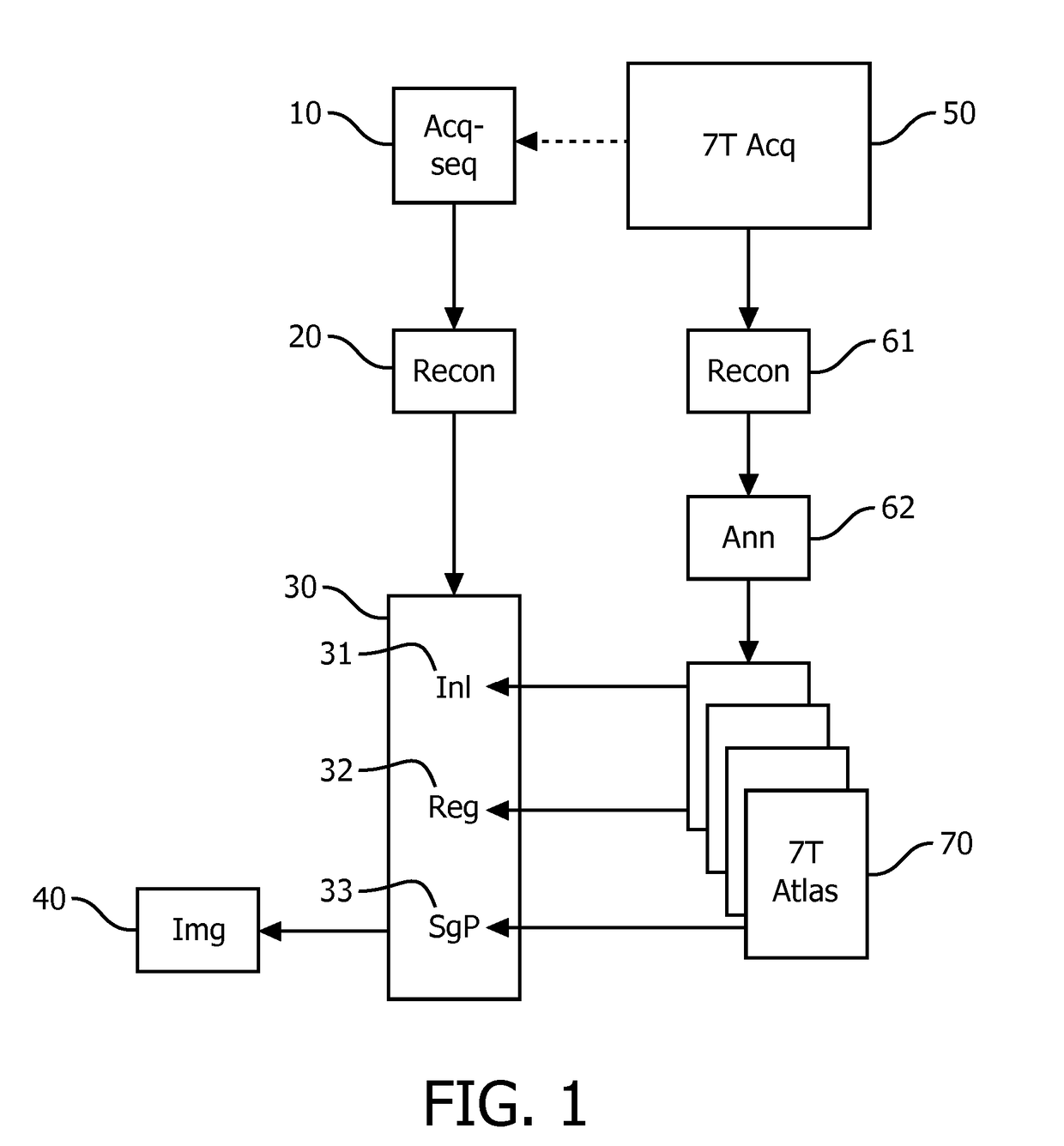
[0042]FIG. 1 shows a schematic representation of a magnetic resonance imaging protocol of the invention. The magnetic resonance signals for the diagnostic magnetic resonance imaging method are acquired in the acquisition segment 10 which is formed by a

[0043]MR acquisition sequence that is translated from the MR acquisition sequence that is employed to generate the training magnetic resonance images from magnetic resonance signal acquired at a higher main magnetic field strength as compared to the field strength at which the magnetic resonance signals for the diagnostic image are acquired. The translation of the MR acquisition sequence at ultra-high (e.g. 7 T) to high (e.g. 3 T) or medium (1.5 T) field strengths achieves that the acquired magnetic resonance signal give rise to a similar image contrast in both the diagnostic and training magnetic resonance images. By way of a reconstruction segment 20, the diagnostic magnetic resonance image is reconstructed from the acquired magnetic...


PUM


Abstract

A magnetic resonance imaging protocol includes an acquisition segment to control an acquisition sequence to acquire magnetic resonance signals at a lower main magnetic field strength. A reconstruction segment controls reconstruction of a diagnostic magnetic resonance image from the magnetic resonance signals at a lower main magnetic field strength. A segmentation segment to control segmentation of a pre-determined image-detail of the diagnostic magnetic resonance image. In the magnetic resonance imaging protocol: the acquisition sequence has a set of imaging parameters that cause the image quality of the diagnostic magnetic resonance to be similar to the image quality of the magnetic resonance training images. The segmentation segment comprises: an initialisation portion which controls (i) access to a set of magnetic resonance training images acquired at main magnetic field of a higher main magnetic field strength (ii) registration of the diagnostic magnetic resonance image to one or more of the magnetic resonance training images and (iii) a segmentation proper applied to the diagnostic image to segment the pre-determined detail from the registered diagnostic magnetic resonance image. The one or more magnetic resonance training images includes an image detail corresponding to the pre-determined image detail in the diagnostic magnetic resonance image. Notably, the magnetic resonance training magnetic resonance images are acquired at a high magnetic field strength at 7 T and its level of detail facilitates the accurate segmentation of notably the hippocampus from diagnostic magnetic resonance images at lower field strength of 3 T. The diagnostic magnetic resonance images are acquired such that they resemble the training magnetic resonance images.

Description

FIELD OF THE INVENTION[0001]The invention pertains to a magnetic resonance imaging protocol for segmentation of an image detail.BACKGROUND OF THE INVENTION[0002]Such a magnetic resonance imaging protocol is known from the paper ‘Automated template-based hippocampal segmentations from MRI: the effects of 1.5 T or 3 T fields strength on accuracy’ by Kate E. Macdonald et al. in Neuroinform. (published on-line on 7 Jan. 2014).[0003]Magnetic imaging protocols in this paper involve acquisition of magnetic resonance signals at different main magnetic field strengths of 1.5 T and 3.0 T. Further, this paper recognises that hippocampal volumetric measures may be useful for Alzheimer's Disease (AD) diagnoses and disease tracking. Because manual segmentation of the hippocampus from magnetic resonance images is labour-intensive, automated segmentation is needed for clinical use. To study its reliability, an automated hippocampal segmentation using hippocampal multi-atlas propagation and segmenta...

Claims


Application Information

Patent Timeline
Patent Type & Authority Applications(United States)
IPC IPC(8): G01R33/56G01R33/561A61B5/055G01R33/44
CPCG01R33/5608G01R33/445A61B5/055G01R33/5617G01R33/5611G01R33/54G01R33/56G01R33/561
Inventor JASINSCHI, RADU SERBANLAMERICHS, RUDOLF MATHIAS JOHANNES NICOLAASBOERNERT, PETER
Owner KONINKLJIJKE PHILIPS NV
Why Eureka
Try Eureka