
Intravascular Plaque Detection in OCT Images

a plaque detection and image technology, applied in image enhancement, instruments, catheters, etc., can solve the problems of insufficient pixels in small windows, difficult to identify which specific textural image characteristic is represented by each of these features, and limited computational complexity of our plaque detection method, so as to reduce the number, reduce processing time, and achieve the effect of sufficient calculation speed

Active Publication Date: 2017-09-07
UNIVERSITY OF MANITOBA
View PDF30 Cites 25 Cited by
  • Summary
  • Abstract
  • Description
  • Claims
  • Application Information

AI Technical Summary

Benefits of technology

The patent describes a method for detecting vascular plaque using only three textural features. This reduces the computation time while maintaining textural information. The method has been validated using histology and photographic images and compares with a more comprehensive set of 26 features. This approach offers an efficient way to detect vascular plaque in real-time. The technical effect of this patent is to provide a reliable and efficient tool for the detection of vascular plaque using a reduced set of textural features and the Fuzzy C-means clustering algorithm.

Problems solved by technology

However, the computational complexity of our plaque detection method was limited by the dimensionality of the feature space.
Even though these textural image features contain information about textural image characteristics of an image, it is difficult to identify which specific textural image characteristic is represented by each of these features [45].
Small windows may not have enough pixels to accurately capture the texture of underlying tissue while too large window may contain tissue of grossly different texture.
To the unaided eye, it will be difficult to differentiate the uniform appearing plaque and the remaining tissue regions from the raw OCT image.

Method used


Image

  • Intravascular Plaque Detection in OCT Images
  • Intravascular Plaque Detection in OCT Images
  • Intravascular Plaque Detection in OCT Images

Examples


Embodiment Construction

[0212]In this work, we obtained vascular tissue samples with atherosclerotic plaque from myocardial infarction prone Watanabe heritable hyperlipidemic rabbits (WHHL rabbits) [16, 17]. Arterial samples were obtained from different locations from three WHHL rabbits aged 10 and 22 months. Arterial segments of tissue starting from the ascending aorta to the external iliac artery were excised from all specimens and subdivided into 20-30 mm long sections. Digital photographs of the luminal surface were taken, and regions of interest were identified prior to measurements. Histology images with oil red O staining were also captured using a Zeiss Axio Observer ZI system (NRC-IBD, Winnipeg, Canada). The oil red O staining emphasizes the lipid content of the tissue, thereby identifying the plaque region. This study was approved by the local animal care committee at the Institute for Biodiagnostics, National Research Council Canada.

[0213]The OCT system used in this work is a catheter based intr...


PUM


Abstract

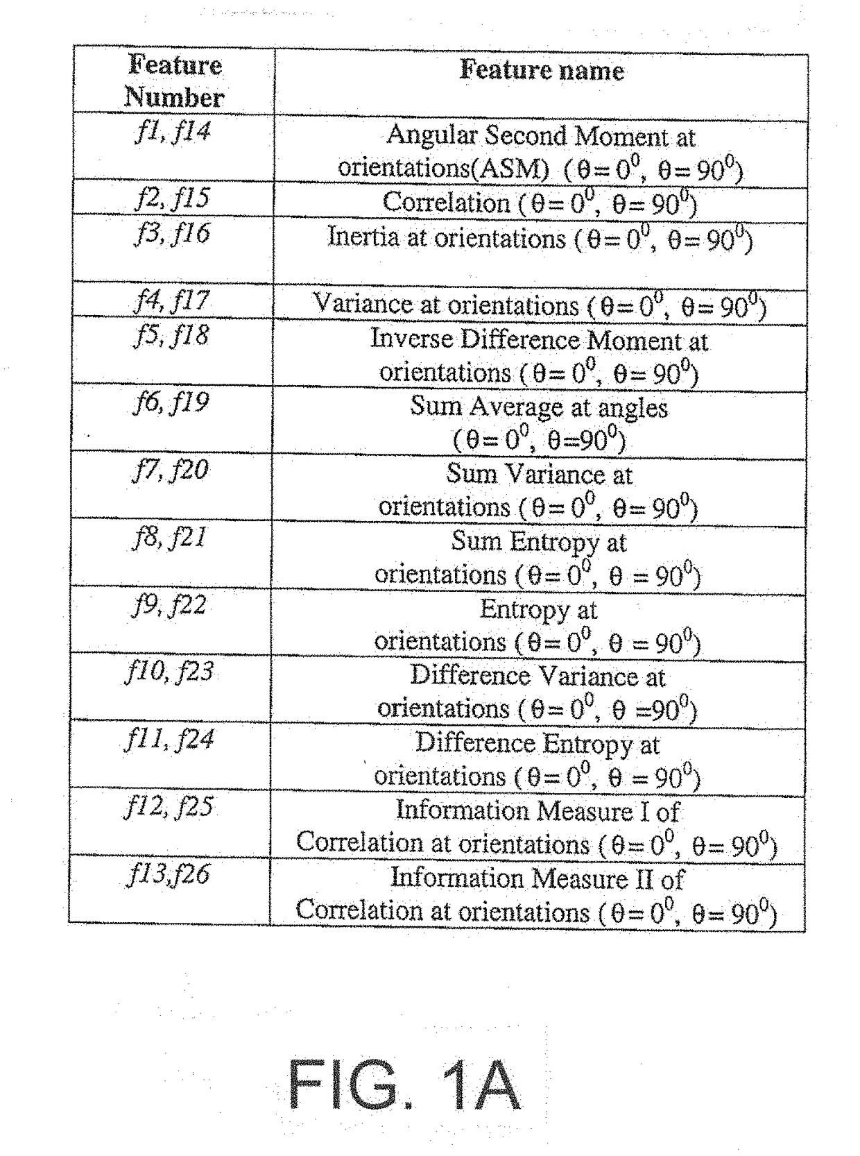
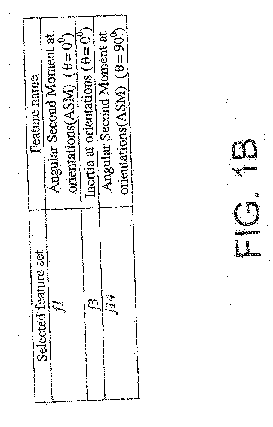
Detection of intravascular plaque in OCT images is carried out by obtaining images of vascular tissue from a vascular component by OCT either in a static mode of a single image or in a dynamic mode where the images are obtained by scanning. The method acts by dividing the OCT image into different regular regions, calculating different texture features for each of the above regions with a reduced set of less than a full set of the 26 Haralick textural features, using a clustering algorithm to segment the image defined by its texture features calculated above into different regions and transforming the segmented image back from its representation using texture features to its space-domain representation. The method uses three or four texture features where the reduced sets can be f1, f 2, and f14 (ASM at 0°, Inertia at 0° and ASM at 90°).

Description

[0001]This application claims the benefit under 35 USC 119 (e) of Provisional Application 62 / 303,767 filed Mar. 4, 2016.[0002]This invention relates to a method of intravascular plaque detection in OCT images.BACKGROUND OF THE INVENTION[0003]Cardiovascular disease is one of the main causes of mortality and morbidity around the globe, and is expected to become the predominant cause of death worldwide [I]. Therefore, it is crucial for medical professionals to be able to detect the conditions that cause cardiovascular disease. The formation of vascular plaque is considered to be the underlying pathology of coronary heart disease as it can accumulate to the point where it blocks arterial blood flow. Many medical imaging methods have been utilized to detect vascular plaque, including: computed tomography (CT) [2-4], intravascular ultrasound (IVUS) [5-8], and magnetic resonance imaging (MRI) [9, 10]. OCT has been proven to be equally effective as high resolution IVUS in detecting calcifie...

Claims


Application Information

Patent Timeline
IPC IPC(8): A61B5/02G06T7/00G06T1/20G06T7/40G06K9/62G06T7/168A61B5/00G06T7/11
CPCA61B5/02007A61B5/0066A61B5/7264G06T7/0012G06T7/11G06T7/40G06T2200/28G06T7/168G06T1/20A61B2576/00G06T2207/10101G06T2207/30024G06T2207/30101G06K9/6218A61B5/6852A61B5/6876A61B5/0084G16H30/40G16H50/20G06V20/698G06V10/763
Inventor PRAKASH, AMMUSHERIF, SHERIF S.
Owner UNIVERSITY OF MANITOBA
Learn More