
Compositions and methods for detection of mycoplasma genitalium

a technology of mycoplasma and genitalium, applied in the field of molecular diagnostics, can solve the problems of inability to readily collect culture samples, inability to detect mycoplasma genitalium, and inability to easily collect culture samples,

Active Publication Date: 2017-11-30
ROCHE MOLECULAR SYST INC
View PDF1 Cites 5 Cited by
  • Summary
  • Abstract
  • Description
  • Claims
  • Application Information

AI Technical Summary

Benefits of technology

The patent describes a method for amplifying specific gene sequences in an efficient and accurate way using a combination of denaturing and incubation at different temperatures. The method involves a one-step setup for both annealing and extension, with the first measurement at a lower temperature to increase inclusion, and the second measurement at a higher temperature to increase specificity. The method has been tested and shown to be effective in amplifying and detecting specific genes in a genomic DNA sample. The results show that the method is highly sensitive and accurate, with Ct values ranging from 15 to 42.

Problems solved by technology

The diagnosis of M. genitalium infection has traditionally been culture of urogenital specimens but culture is insensitive and slow and may take up to 6 months to recover the organism.
The fact that M. genitalium is a slow-growing organism, culture is not readily available, slow and insensitive, NAATs are the preferred method for M. genitalium detection.

Method used


Image

  • Compositions and methods for detection of mycoplasma genitalium
  • Compositions and methods for detection of mycoplasma genitalium
  • Compositions and methods for detection of mycoplasma genitalium

Examples


example 1

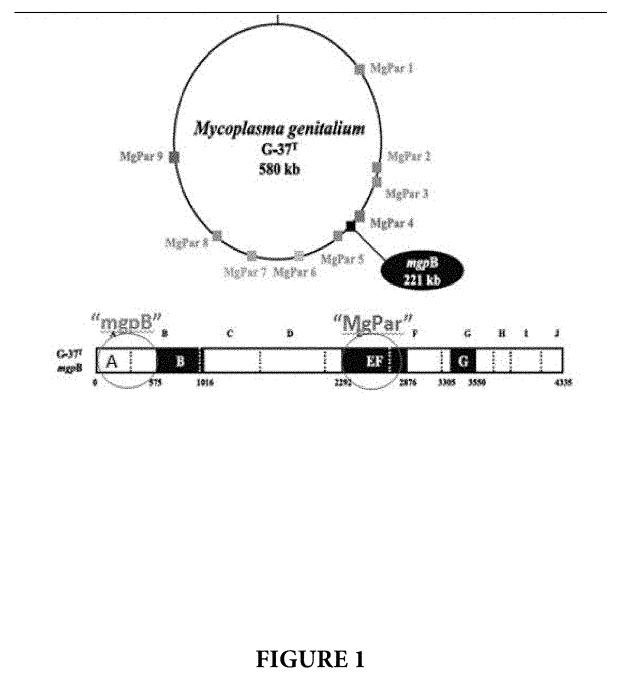
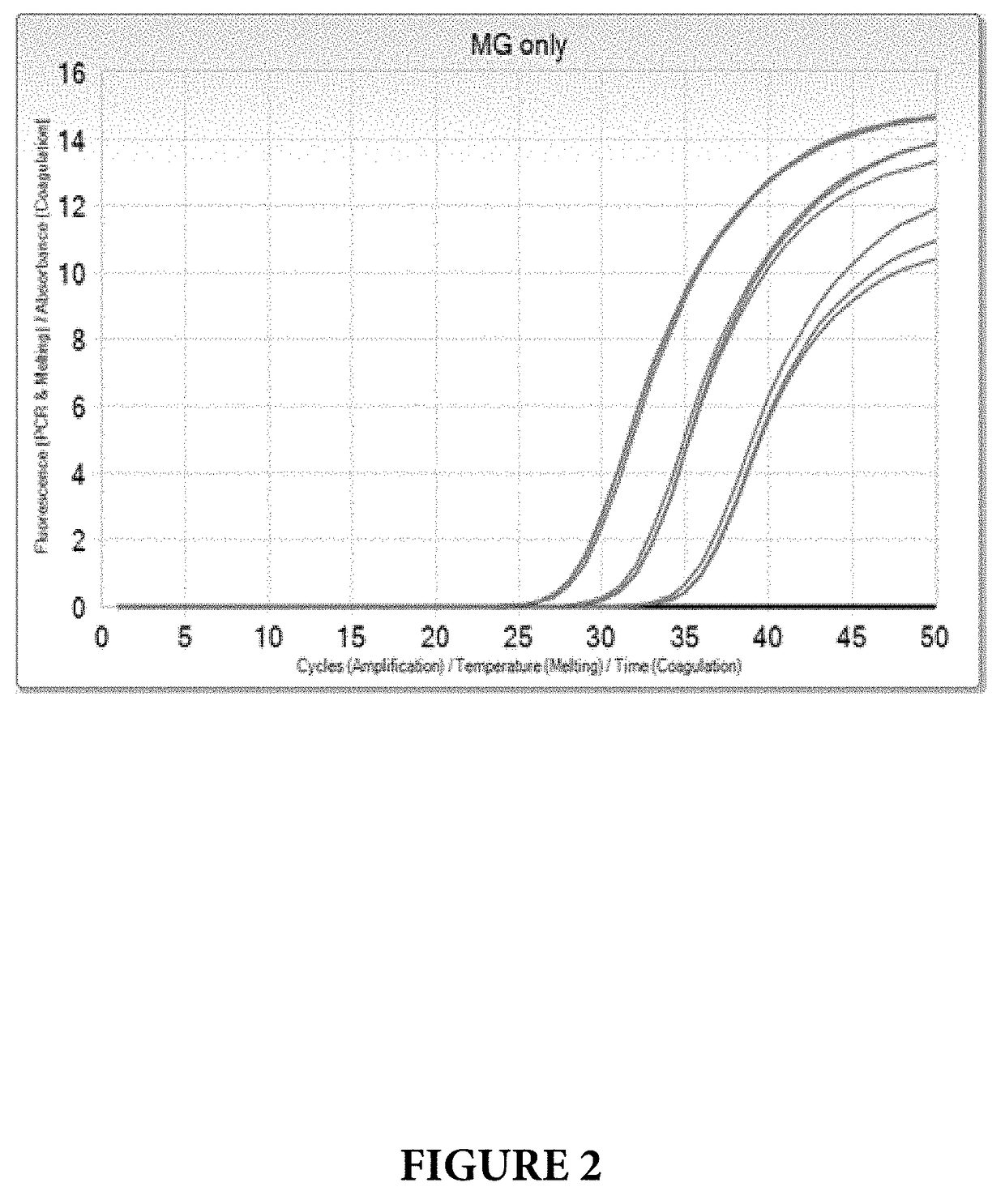
[0091]Target selection for MG was the result of a comprehensive search of the public sequence database. Multiple methods were used to search for Mycoplasma genitalium sequences and its nearest neighbor, Mycoplasma pneumoniae, to design for assay exclusivity. The MG targets selected had the most coverage in the public database (mgpB 120 sequences, MgPar 5 whole genome assemblies). MG has a small compact genome, which limited regions to target for NAT design. The mgpB surface protein gene (called the MgPar target) was selected for the assay due to its multi-copy number. Surface proteins are known to undergo recombination, which can result in a failed assay. To mitigate the associated risks, a dual target approach was implemented using a well conserved target (called the mgpB target).

[0092]The MgPa operon codes three genes: mgpA, mgpB and mgpC (black circle labeled as mgpB 221 kb in FIG. 1). mgpB (4335 bp) is present in the complete operon but there are nine partial repeats (MgPar's) o...

example 2

[0096]The amplification and detection of the MG mgpB and the MG MgPar genes was performed as described in Example 1 with the exception that genomic template DNA for Trichomonas vaginalis (TV) was included in the PCR assay together with primers and probes that can amplify and detect TV. In this experiment, primers and probes, disclosed in U.S. Provisional Application No. 62 / 342,600, that hybridize to the 5.8s rRNA gene were used.

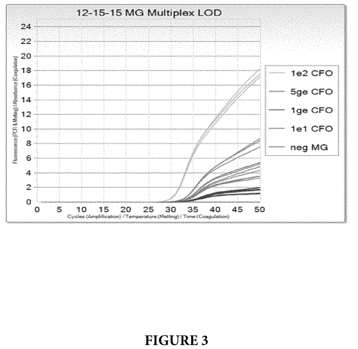
[0097]MG Limit of Detection (LOD) was tested at 100, 10, 5 and 1 genomic equivalent concentrations per PCR reaction (ge / PCR), in a co-amplification with internal control standard and TV at 10 ge / PCR. The results are shown on FIGS. 3 and 4. All levels of MG were detected with no dropouts, and MG LOD was determined to be <1 ge / PCR.

[0098]While the foregoing invention has been described in some detail for purposes of clarity and understanding, it will be clear to one skilled in the art from a reading of this disclosure that various changes in form and detail can ...


PUM

PropertyMeasurementUnit
temperatureaaaaaaaaaa

Abstract

Methods for the rapid detection of the presence or absence of Mycoplasma genitalium (MG) in a biological or non-biological sample are described. The methods can include performing an amplifying step, a hybridizing step, and a detecting step. Furthermore, primers, probes targeting the target MG gene, along with kits are provided that are designed for the detection of MG.

Description

CROSS REFERENCE TO RELATED APPLICATIONS[0001]This application claims the benefit of priority of U.S. Provisional Patent Application Ser. No. 62 / 342,519, filed May 27, 2016, which is incorporated herein by reference in its entirety.REFERENCE TO SEQUENCE LISTING[0002]This application contains a Sequence Listing submitted as an electronic text file named “33594_US1_Sequence_Listing.txt”, having a size in bytes of 30 kb, and created on Apr. 17, 2017. The information contained in this electronic file is hereby incorporated by reference in its entirety pursuant to 37 CFR §1.52(e)(5).FIELD OF THE INVENTION[0003]The present disclosure relates to the field of molecular diagnostics, and more particularly to detection of Mycoplasma genitalium. BACKGROUND OF THE INVENTION[0004]Mycoplasma genitalium (MG) is a gram-negative bacterium that lacks a cell wall and lives on the surface and in the epithelial cells of the urinary and genital tracts of men and women. M. genitalium is a common cause of n...

Claims


Application Information

Patent Timeline
Patent Type & Authority Applications(United States)
IPC IPC(8): C12Q1/68
CPCC12Q2600/158C12Q1/689C12Q1/686C12Q2537/143C12Q2565/1025C12Q2600/16
Inventor HARRIS, JODYMANGIPUDI, KALYANI
Owner ROCHE MOLECULAR SYST INC
Learn More