Endoscopic diagnosis apparatus, image processing method, program, and recording medium
- Summary
- Abstract
- Description
- Claims
- Application Information
AI Technical Summary
Benefits of technology
Problems solved by technology
Method used
Image
Examples
first embodiment
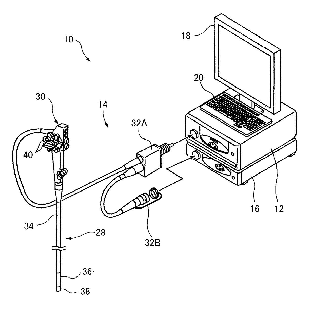
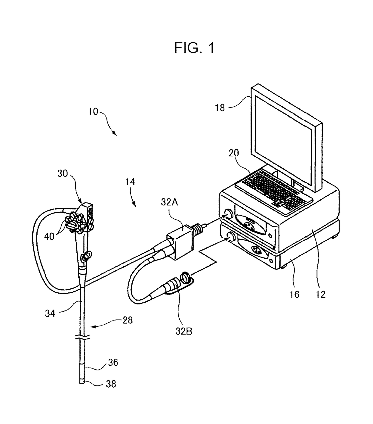
[0084]First, a description will be given of, as a first embodiment, the case of measuring the actual size of a subject in an endoscopic image by using the hood attached to the distal end portion of the endoscope 14.
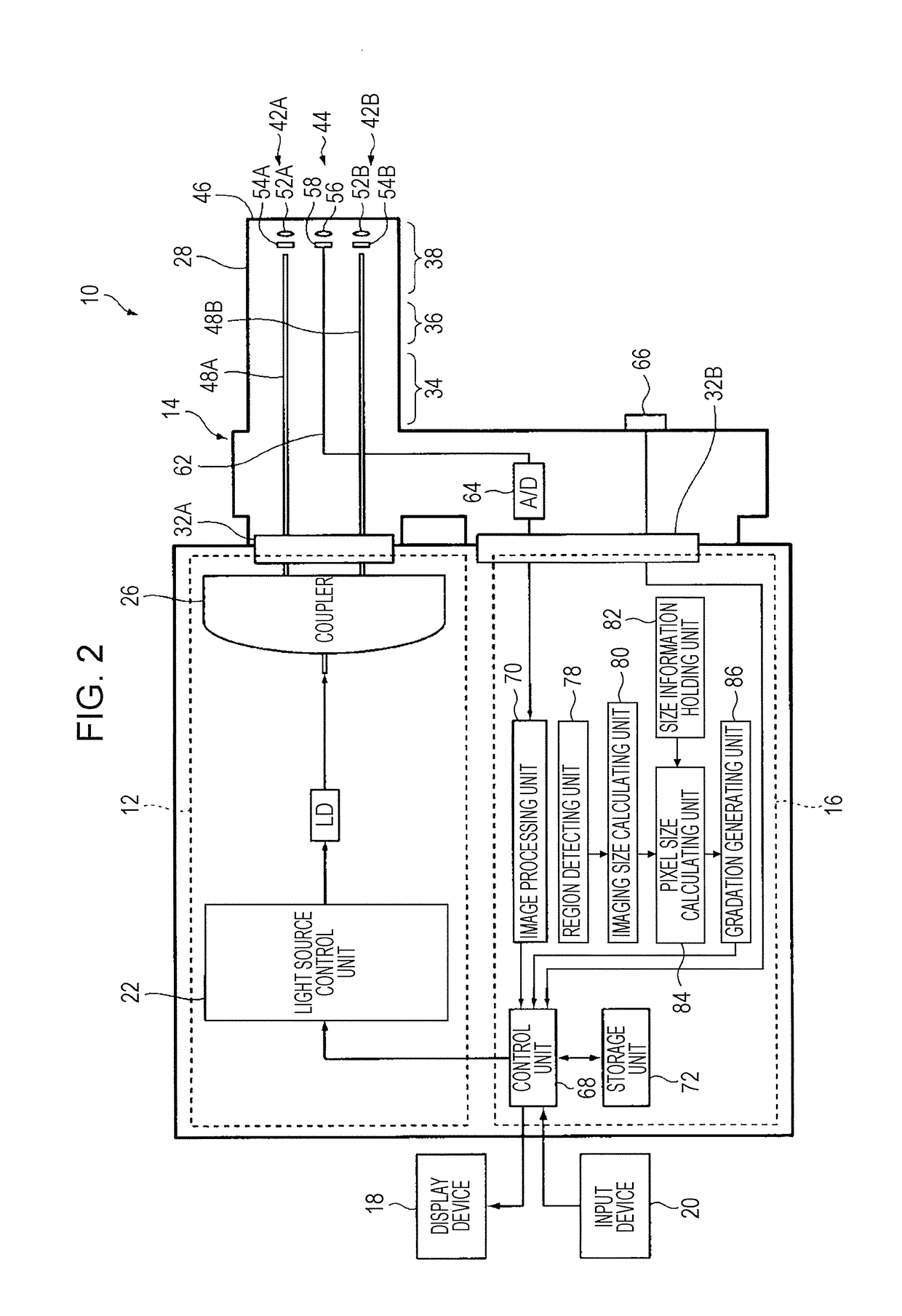
[0085]First, an operator of the endoscopic diagnosis apparatus 10 inputs through the input device 20 information of the actual size of the other opening portion of the hood attached to the distal end portion of the endoscope 14. The information of the actual size of the other opening portion of the hood is held by the size information holding unit 82.
[0086]Subsequently, the operator inserts the endoscope 14 whose distal end portion is attached with the transparent hood into a subject and moves the endoscope 14 to a region of interest while checking an endoscopic image displayed on the display device 18, and then the other opening portion of the hood comes in contact with a surface of the region of interest of the subject, as illustrated in FIG. 6A.
[0087]Subsequently, the ...
second embodiment
[0097]Next, a description will be given of, as a second embodiment, the case of measuring the actual size of a subject in an endoscopic image by using a surgical instrument.
[0098]As in the case of the first embodiment, the operator inputs through the input device 20 information of the actual size of a tip portion of a surgical instrument extending outward from the forceps outlet 74 at the distal end portion of the endoscope 14. The information is held by the size information holding unit 82.
[0099]Subsequently, the operator inserts the endoscope 14 into a subject and moves the endoscope 14 to a region of interest while checking an endoscopic image displayed on the display device 18. Subsequently, the operator inserts the surgical instrument from the forceps inlet of the endoscope 14 so that the surgical instrument is extended outward from the forceps outlet 74 at the distal end portion of the endoscope 14, and the tip portion of the surgical instrument comes in contact with a surface...
third embodiment
[0110]Next, a description will be given of, as a third embodiment, the case of measuring the actual length of a subject in an endoscopic image by using a water jet.
[0111]As in the case of the first embodiment, the operator inputs through the input device 20 information of the actual size of the ejection opening from which a water jet is ejected at the distal end portion of the endoscope 14. The information is held by the size information holding unit 82.
[0112]Subsequently, the operator inserts the endoscope 14 into a subject and moves the endoscope 14 to a region of interest while checking an endoscopic image displayed on the display device 18. Subsequently, a water jet is ejected from the ejection opening at the distal end portion of the endoscope 14 to a surface of the region of interest of the subject, as illustrated in FIG. 9.
[0113]Subsequently, the operator presses a button or the like located in the operation section 30 of the endoscope 14 to input an instruction to start dete...
PUM
 Abstract
Description
Claims
Application Information
 - R&D
- Intellectual Property
- Life Sciences
- Materials
- Tech Scout
- Unparalleled Data Quality
- Higher Quality Content
- 60% Fewer Hallucinations
Browse by: Latest US Patents, China's latest patents, Technical Efficacy Thesaurus, Application Domain, Technology Topic, Popular Technical Reports.
© 2025 PatSnap. All rights reserved.Legal|Privacy policy|Modern Slavery Act Transparency Statement|Sitemap|About US| Contact US: help@patsnap.com


