

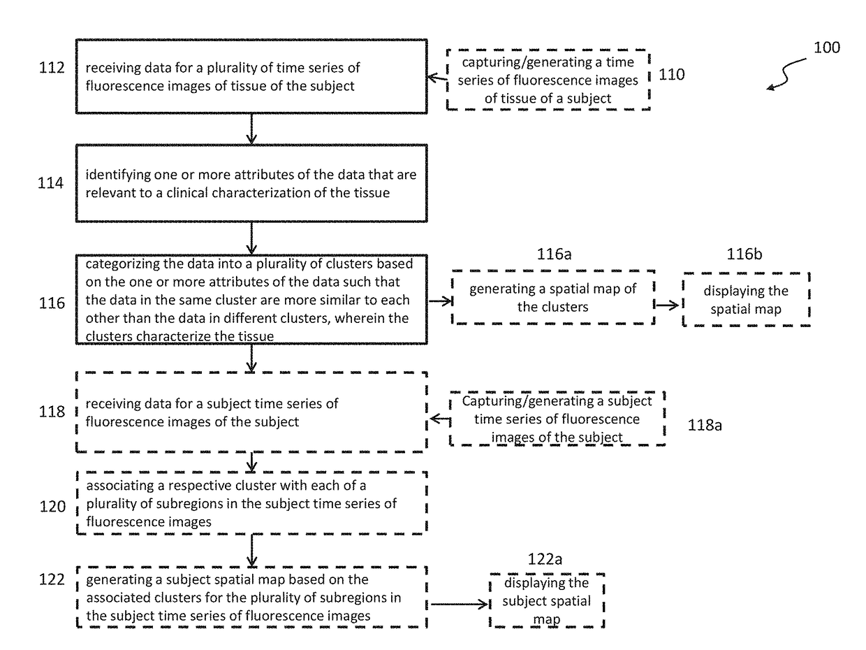
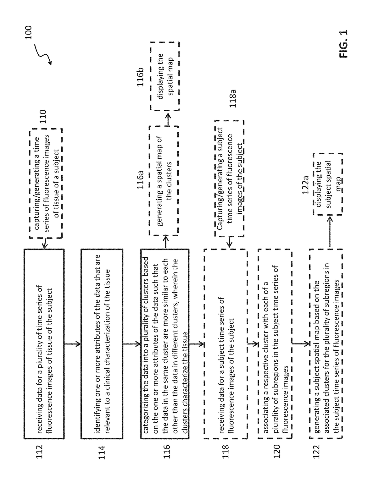
Methods and systems for characterizing tissue of a subject utilizing a machine learning
a machine learning and tissue technology, applied in the field of imaging, can solve the problems of insufficient quality visual evaluation of such images, limited visual evaluation, and inability to support a standardized protocol for assessing blood flow and/or tissue perfusion, and achieve the effect of facilitating the acquisition and generation of visual representations
- Summary
- Abstract
- Description
- Claims
- Application Information
AI Technical Summary
Benefits of technology
Problems solved by technology
Method used
Image

Examples
examples
Application of the Methods and Systems in Wound Management
[0183]One challenge in wound management, such as chronic wound management, is that the medical condition or nature of a wound can be viewed differently among clinicians. Conventional techniques may provide information about the wound's pathological history, but fail to provide reliable indicators of viability and / or restorative potential, e.g., whether wound and / or periwound is likely to develop complications, is capable of healing, or how healing progresses (e.g., time to achieve an acceptable healing stage). Furthermore, wounds exist where no pathology is demonstrable by conventional diagnostic techniques. Various embodiments of the methods and systems described herein facilitate producing a consistent representation (not subjective to biases of perception) of the state of a particular tissue region (e.g., wound, periwound), and thus facilitate a more accurate subsequent assessment and formulation of care strategies (e.g., ...
PUM
 Abstract
Description
Claims
Application Information