Myocardial excitation complementation/visualization apparatus and myocardial excitation detection apparatus

a detection apparatus and myocardial excitation technology, applied in the field of myocardial excitation complementation/visualization apparatus and myocardial excitation detection apparatus, can solve the problems of increasing the possibility of brain infarction or the like, increasing the computational amount of computation process, and reducing the computational amount so as to improve the accuracy of detecting the position of excitation in the myocardium, enhance the accuracy of generating visualized data, and reduce the computational amoun

Active Publication Date: 2018-03-15
NAT UNIV CORP SHIGA UNIV OF MEDICAL SCI +3
View PDF0 Cites 4 Cited by
  • Summary
  • Abstract
  • Description
  • Claims
  • Application Information

AI Technical Summary

Benefits of technology

The patent describes a method for visualizing the state of excitation in the heart using intracardiac electrocardiograms. The method involves preparing waveforms from the electrocardiograms, generating phase portraits, and using a pseudo action potential transform to reduce the amount of computation required. This results in the ability to continuously prepare visualized data and display it in real-time. The method also improves the accuracy of detecting the position of excitation in the heart. Overall, the method provides a novel way to visualize and analyze the heart's electrical activity.

Problems solved by technology

Generally, atrial fibrillation means an arrhythmia in which the atrium of the heart has a convulsive seizure, and the heart cannot operate in the original and correct way.
When the atrial fibrillation occurs, the blood stagnates in the atrium, and thrombus is prone to be formed, thereby increasing the possibility of occurrence of brain infarction or the like.
In the Hilbert transform, however, a fast Fourier transform (FFT) and an inverse fast Fourier transform (IFFT) are performed in one computation, and therefore throughput of the computation process is huge.
Because of the huge computation amount, under present circumstances, the application of the prior art using the Hilbert transform is limited to a use in an off-line mode.
In the prior art, as described above, it is difficult to continuously prepare visualized data with respect to an intracardiac electrocardiogram which is recorded by a cardiac catheter, and display the data in real time.

Method used


Image

  • Myocardial excitation complementation/visualization apparatus and myocardial excitation detection apparatus
  • Myocardial excitation complementation/visualization apparatus and myocardial excitation detection apparatus
  • Myocardial excitation complementation/visualization apparatus and myocardial excitation detection apparatus

Examples


embodiment 1

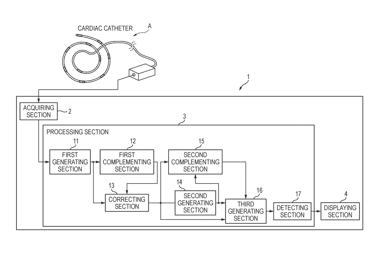
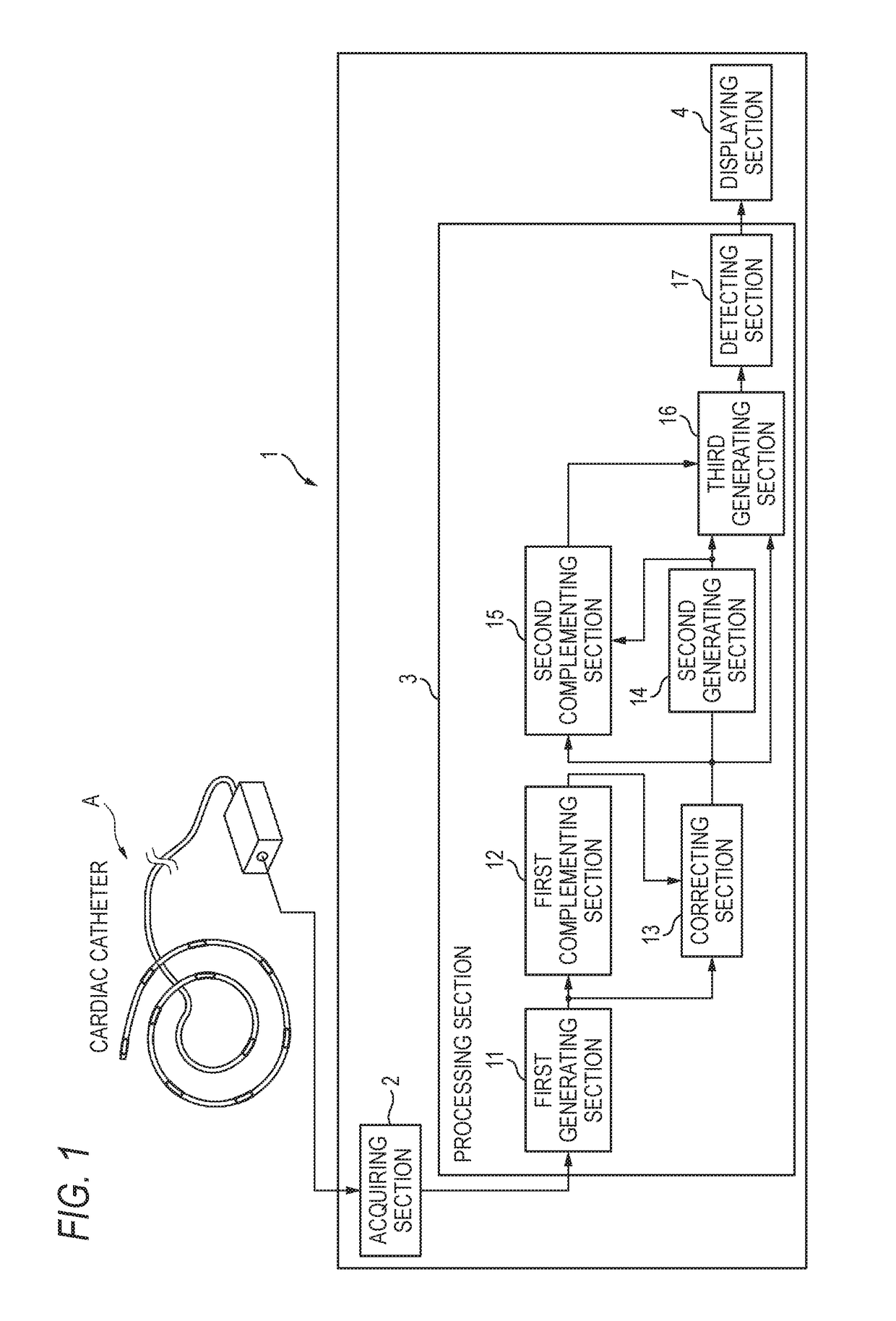
[0044]As shown in FIG. 1, a myocardial excitation complementation / visualization apparatus 1 of Embodiment 1 includes an acquiring section 2, a processing section 3, and a displaying section 4. For example, the myocardial excitation complementation / visualization apparatus 1 is used as an apparatus for performing one function of a catheter inspection apparatus.

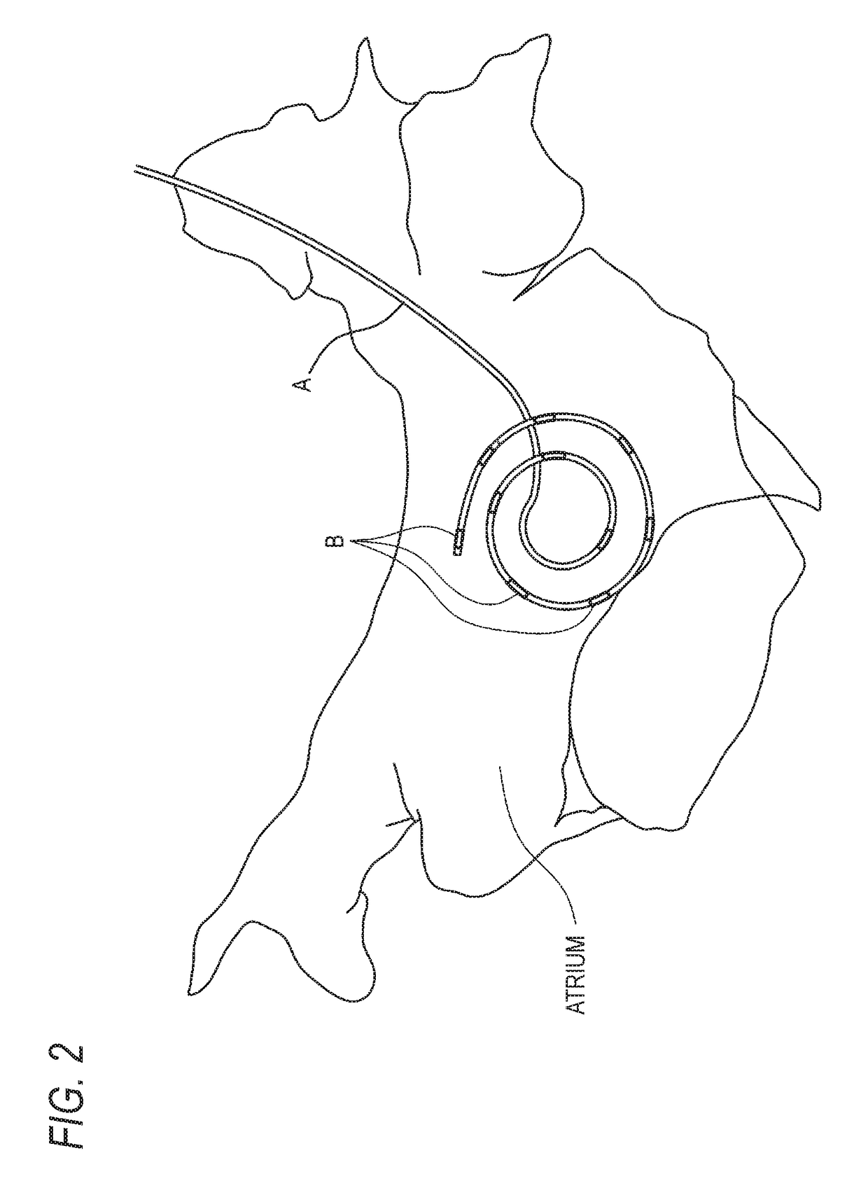
[0045]The acquiring section 2 acquires an intracardiac electrocardiogram of a subject which is recorded by a recording unit A (e.g., a cardiac catheter) having a plurality of electrodes.

[0046]The processing section 3 performs a computation for visualizing the state of myocardial excitation of the subject, on the intracardiac electrocardiogram which is acquired by the acquiring section 2. The processing section 3 includes a first generating section 11, a first complementing section 12, a correcting section 13, a second generating section 14, a second complementing section 15, a third generating section 16, and a detecting section...

embodiment 2

[0096]Next, Embodiment 2 will be described. Hereinafter, components which are identical with those of Embodiment 1 are denoted by the same reference numerals, and their description is omitted.

[0097]As shown in FIG. 20, a myocardial excitation complementation / visualization apparatus 100 of Embodiment 2 includes the acquiring section 2, a processing section 3A, a storage section 110, and the displaying section 4. The processing section 3A includes a first generating section 111, the first complementing section 12, the second generating section 14, the second complementing section 15, the third generating section 16, and the detecting section 17.

[0098]The storage section 110 stores a plurality of action potential unit waveforms 120 which are as shown in, for example, FIG. 21, and which are previously generated. The action potential unit waveforms 120 are obtained by applying a temporal moving averaging process on an action potential waveform in the human atrial muscle under structural ...


PUM


Abstract

A myocardial excitation complementation / visualization apparatus includes an acquiring section that acquires intracardiac electrocardiograms of a subject, the intracardiac electrocardiograms being recorded by a recording unit having a plurality of electrodes, a processing section that performs a computation for completing and visualizing a state of excitation in a myocardium of the subject based on the intracardiac electrocardiograms, and a displaying section that displays the state of excitation in the myocardium of the subject based on an output of the processing section. The processing section includes a first generating section, a correcting section, a second generating section, and a third generating section. The displaying section displays a change of the state of excitation in the myocardium of the subject based on the visualized data.

Description

BACKGROUNDTechnical Field[0001]The present invention relates to a myocardial excitation complementation / visualization apparatus for visualizing the state of excitation in the myocardium, and also to a myocardial excitation detection apparatus for detecting excitation in the myocardium.Background Art[0002]Generally, atrial fibrillation means an arrhythmia in which the atrium of the heart has a convulsive seizure, and the heart cannot operate in the original and correct way. When the atrial fibrillation occurs, the blood stagnates in the atrium, and thrombus is prone to be formed, thereby increasing the possibility of occurrence of brain infarction or the like.[0003]Conventionally, it is known that, when an arrhythmia such as atrial fibrillation occurs, a treatment is performed by selectively applying ablation to an abnormal portion which causes the arrhythmia, by using a cardiac catheter. In the treatment, it is important to correctly identify the location where ablation is to be per...

Claims


Application Information

Patent Timeline
IPC IPC(8): A61B5/046A61B5/0408A61B5/0456A61B5/00A61B5/361A61B5/296A61B5/352
CPCA61B5/046A61B5/0408A61B5/0456A61B5/743A61B5/04023A61B5/02A61B5/0006A61B18/1492A61B5/318A61B5/322A61B5/352A61B5/25A61B5/361A61B5/349
Inventor ASHIHARA, TAKASHITAKIZAWA, KOJINISHIHARA, TATSUOSUZUKI, NOBUHIROIWANAGA, YUUHOOTA, AKIOTSUCHIYA, TAKESHINAKAZAWA, KAZUOINADA, SHIN
Owner NAT UNIV CORP SHIGA UNIV OF MEDICAL SCI
Why Eureka
Try Eureka