Systems and methods for calibrating a nuclear medicine imaging system

a nuclear medicine and imaging system technology, applied in the field of medical imaging systems, can solve the problems of time-consuming and difficult approach, inability to calibrate multiple isotopes (having different energy peaks) after the system is assembled, and inability to achieve the effect of multiple isotopes

Active Publication Date: 2018-04-26
GENERAL ELECTRIC CO
View PDF4 Cites 4 Cited by
  • Summary
  • Abstract
  • Description
  • Claims
  • Application Information

AI Technical Summary

Benefits of technology

[0005]In one embodiment, a method comprises: detecting, with a plurality of detectors, photons emitted by a calibration source comprising a radioactive line source and a fluorescence source, while pivoting one or more detectors of the plurality of detectors; and calibrating, with a processor communicatively coupled to the plurality of detectors, each detector of the plurality of detectors based on energy measurements of the detected photons. In this way, a two-point energy calibration of detectors can be performed with a single isotope, and without removing or adjusting a collimator attached to the detector.

Problems solved by technology

In some collimated detector systems, calibration with multiple isotopes (having different energy peaks) is not practical after the system is assembled.
Such an approach is time-consuming and difficult, as the detectors must be disassembled and multiple calibrations must be performed.

Method used


Image

  • Systems and methods for calibrating a nuclear medicine imaging system
  • Systems and methods for calibrating a nuclear medicine imaging system
  • Systems and methods for calibrating a nuclear medicine imaging system

Examples


Embodiment Construction

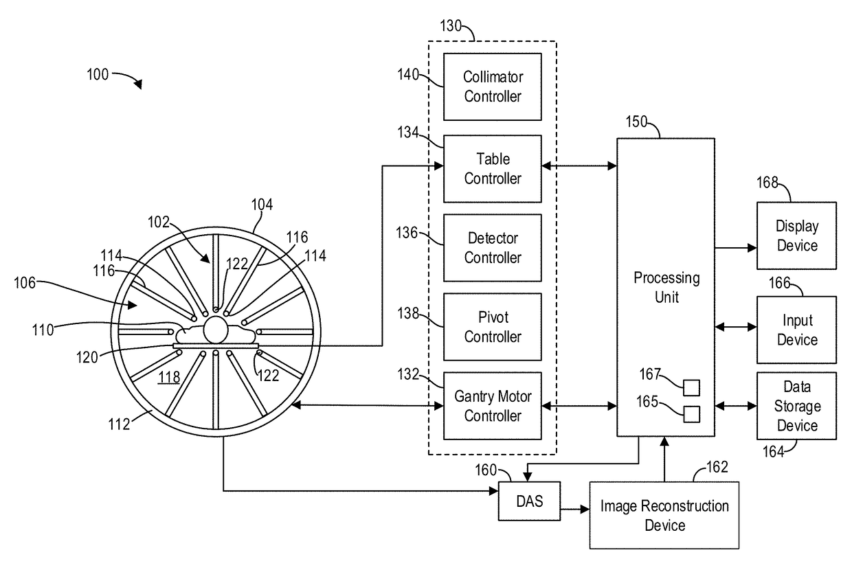
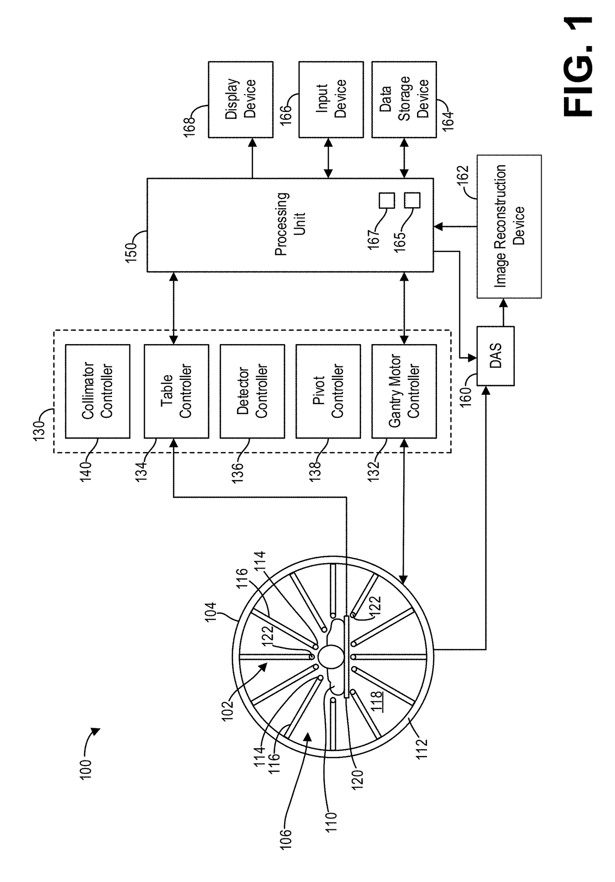
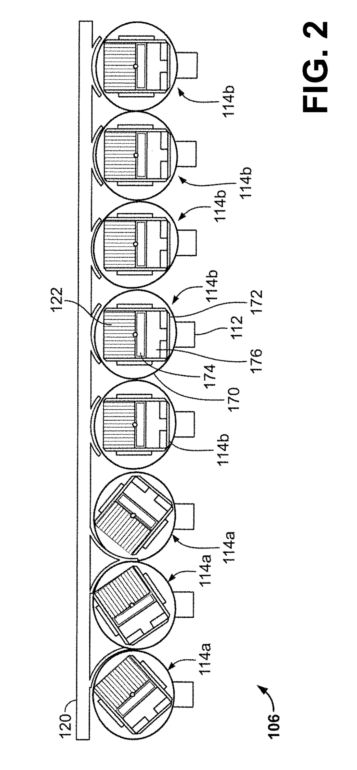
[0021]The following description relates to various embodiments of nuclear medicine imaging systems. In particular, systems and methods are provided for a calibration source for a nuclear medicine (NM) imaging system that enables calibration of detectors of the NM imaging system at multiple energy peaks (e.g., two-peak energy calibration) using a single isotope. An imaging system, such as the imaging system depicted in FIG. 1, may include systems for controlling the movement of a plurality of imaging detectors to position the imaging detectors to acquire image data. For example, in various embodiments a Nuclear Medicine (NM) imaging system with an array of detector heads that are individually and independently movable is provided, as depicted in FIG. 2. In some embodiments, one or more of the heads are capable of a plurality of types of movement, such as rotation and linear motion. For example, the detector heads may be configured to be positioned adjacent or proximate to a subject a...


PUM


Abstract

Methods and systems are provided for calibrating a nuclear medicine imaging system. In one embodiment, a method comprises: detecting, with a plurality of detectors, photons emitted by a calibration source comprising a radioactive line source and a fluorescence source, while pivoting one or more detectors of the plurality of detectors; and calibrating, with a processor communicatively coupled to the plurality of detectors, each detector of the plurality of detectors based on energy measurements of the detected photons. In this way, a two-point energy calibration of detectors can be performed with a single isotope, and without removing or adjusting a collimator attached to the detector.

Description

FIELD[0001]Embodiments of the subject matter disclosed herein relate to medical imaging systems, and more particularly, to calibrating a nuclear medicine (NM) imaging system.BACKGROUND[0002]In nuclear medicine imaging, systems with multiple detectors or detector heads may be used to image a subject. For example, the detectors may be positioned adjacent to the subject to acquire nuclear medicine imaging data (e.g., radioactivity), which is used to generate a three-dimensional (3D) image of the subject. In a specific example, Single Photon Emission Computed Tomography (SPECT) systems may have moving detector heads, such as gamma cameras, positioned to focus on a region of interest. One or more of the gamma cameras may be moved (for example, rotated) to different angular positions to acquire image data. The acquired image data may then be used to generate 3D images.[0003]In single photon imaging systems, such as planar or SPECT imaging systems, collimators may be placed in front of a s...

Claims


Application Information

Patent Timeline
Patent Type & Authority Applications(United States)
IPC IPC(8): A61B6/00
CPCA61B6/582A61B6/037A61B6/06A61B6/545A61B6/585
Inventor LEVY, MOSHEBOUHNIK, JEAN-PAULGROBSHTEIN, YARIVAMISAR, GILHEFETZ, YARON
Owner GENERAL ELECTRIC CO
Why Eureka
Try Eureka