Cell observation apparatus

Inactive Publication Date: 2018-08-30
SHIMADZU CORP
View PDF3 Cites 1 Cited by
  • Summary
  • Abstract
  • Description
  • Claims
  • Application Information

AI Technical Summary

Benefits of technology

[0028]According to the above configuration, it is possible to adjust the contrast of the phase image colored in accordance with the default color scale or the color scale set by the operator to be optimized without the operator's manual adjustment of the contrast. As a result, it is possible to efficiently observe the cells by eliminating a labor of the operator.
[0029]According to the cell observation apparatus acco

Problems solved by technology

In general, biological cells are colorless and transparent and difficult to observe with a normal optical microscope.
For this reason, a contrast between a cell part to

Method used


Image

  • Cell observation apparatus
  • Cell observation apparatus
  • Cell observation apparatus

Examples


Example

[0034]Hereinafter, an embodiment of a cell observation apparatus according to the present invention will be described with reference to the accompanying drawings.

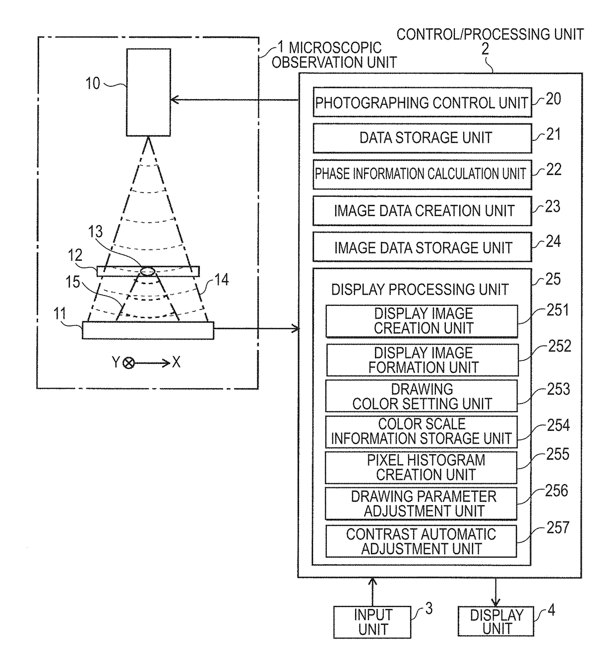
[0035]FIG. 1 is a schematic configuration diagram of the cell observation apparatus according to the present embodiment.

[0036]The cell observation apparatus according to the present embodiment includes a microscopic observation unit 1, a control / processing unit 2, an input unit 3 and a display unit 4 as a user interface.

[0037]The microscopic observation unit 1 is an in-line holographic microscope (IHM), and includes a light source unit 10 including a laser diode and the like and an image sensor 11, and a cell culture plate 12 containing a cell 13 to be observed is placed between the light source unit 10 and the image sensor 11.

[0038]The control / processing unit 2 controls an operation of the microscopic observation unit 1 and processes the data acquired by the microscopic observation unit 1, and includes a photographing cont...


PUM


Abstract

A display image formation unit reads image data according to a designated condition from an image data storage unit to form a phase image, a drawing color setting unit colorizes a phase image according to a color scale in which a phase value in the vicinity of 0 is associated with black, a phase value in a positive direction is associated with a predetermined color other than black, and a phase value in a negative direction is associated with another color other than black. In an area where no cell exists on a medium, the phase value becomes almost 0, so that the background area of the cell on the colorized phase image becomes almost black and the cell is depicted in a color other than black.

Description

FIELD[0001]The present invention relates to a cell observation apparatus for observing biological cells, and more particularly, to a cell observation apparatus for observing biological cells using a phase contrast microscopic image obtained by a phase contrast microscope, a phase image created on the basis of a hologram by a digital holographic microscope, and the like.BACKGROUND ART[0002]In the field of regenerative medicine, studies using pluripotent stem cells such as iPS cells and ES cells have been actively conducted in recent years. In general, biological cells are colorless and transparent and difficult to observe with a normal optical microscope. Therefore, a phase contrast microscope is widely used to observe biological cells (see, Patent Literature 1 and the like). For the phase contrast microscope, when illumination light passes through an observation target, a two-dimensional observation image can be created with contrast of light and dark, on the basis of a difference b...

Claims


Application Information

Patent Timeline
IPC IPC(8): G06T5/00G06T5/40G06T11/00G02B21/36
CPCG06T5/007G06T5/40G06T11/001G02B21/368G02B21/365G06T2207/10056G06T2207/10061G06T7/11G02B21/14G06T2207/30024G02B21/0052G03H1/0005G03H2001/005
Inventor AOI, YUSUKEIWAO, YOSHIHIKO
Owner SHIMADZU CORP
Why Eureka
Try Eureka