
Eureka
For R&D, Eureka makes reading and utilizing patents & technical documents easy.
Eureka AIR
Designed for self-driven R&D workflows. Generate viable solutions, solve complex R&D challenges, empower your innovation with AI.

Eureka Materials
Designed for material experts only. Revolutionize your material R&D, from search, analyze, to developing new materials.

TechResearch
Generate reliable direction feasibility study reports for your R&D in just a few steps.

TechSeek
Discover and master advanced knowledge NOW. Basics, ideas, possibilities, all at once.

TechMind
As an expert in R&D Theories, TechMind can generates customized viable solutions instantly.

TechRisk
Analyze your overall solution with one click, know your potential R&D risks in advance.

TechMonitor
Get weekly tech updates, stay abreast of the latest tech innovations and key insights.
Compositions and methods to improve nanoparticle distribution within the brain interstitium
- Summary
- Abstract
- Description
- Claims
- Application Information
AI Technical Summary
Benefits of technology
Problems solved by technology
Method used
Image

Examples
example 1
f Coating Particles with Non-Adhesive Coating
[0141]Materials and Methods
[0142]i. Nanoparticle Preparation and Characterization
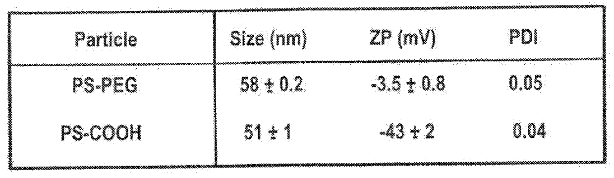
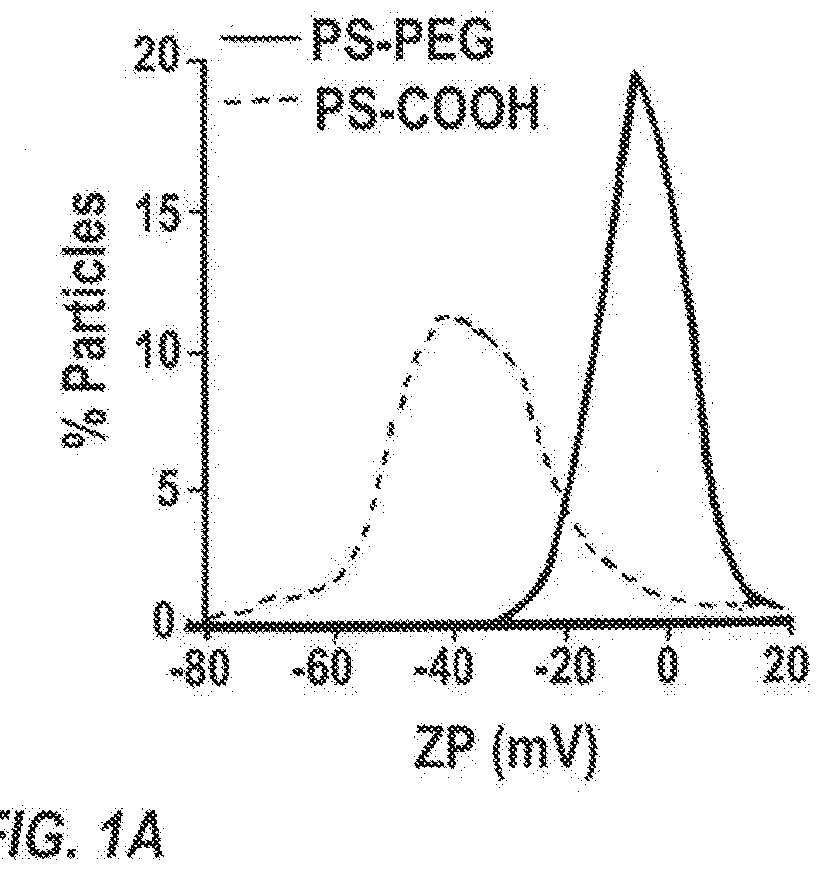
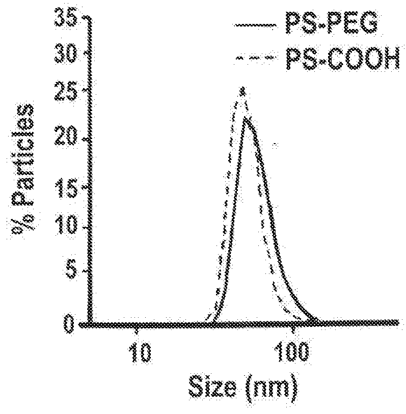
[0143]40-nm dark red fluorescent carboxylated polystyrene microspheres (PS-COOH) (Life Technologies, Grand Island, N.Y.) were modified by conjugating a dense layer of 5 kDa methoxy-PEG-amine (Creative PEGworks, Winston Salem, N.C.), onto the surface, according to a previously published protocol (Nance, E. A., et al., Sci Transl Med, 2012, 4(149), 149ra119), to obtain densely PEGylated polystyrene nanoparticles (PS-PEG). PLGA (75:25) (MW: 15 kDa; Jinan Daigang Biomaterials Co. Ltd., Jinan, China) and PLGA-PEG (75:25) (25 wt % PEG; Jinan Daigang Biomaterials Co. Ltd., Jinan, China) nanoparticles were formulated using the single emulsion process according to a previously published protocol (Nance, E., et al., ACS Nano, 2014, 8(10), 10655-10664). Briefly, PLGA-PEG and PLGA polymer were fluorescently labeled with AlexFluor 647 and AlexaFluor 555 cadaverine dye (Mo...
example 2
f Different Concentrations of Infusate Composition
[0159]Materials and Methods
[0160]i. Ex Vivo Characterization of Brain Pore Sizes
[0161]Brains from female CF-1 mice were harvested and multiple particle tracking was conducted on nanoparticles injected into 1.5 mm thick brain slices according to a slightly modified protocol of Nance, et al., Sci Transl Med, 2012, 4(149), 149ra119. Briefly, the harvested rodent brain was rinsed in chilled artificial spinal fluid and sliced at 1.5 mm intervals using a Zivic mouse brain mold (Zivic Instruments, Pittsburgh, Pa.). Individual brain slices were immersed in infusate solutions (water, 0.9% saline, 3% saline, 10% mannitol, or 25% mannitol) for 5 minutes. Brain slices were removed and mounted on a custom made well and 0.5 μL of fluorescently labeled PS-PEG nanoparticles were injected into the cortex. A coverslip was glued on top of the specimen to prevent bulk flow in the tissue. The particle trajectories were recorded as 20 second movies at an ...
PUM
| Property | Measurement | Unit |
|---|---|---|
| Fraction | aaaaa | aaaaa |
| Fraction | aaaaa | aaaaa |
| Fraction | aaaaa | aaaaa |
Abstract
Description
Claims
Application Information
 - R&D Engineer
- R&D Manager
- IP Professional
- Industry Leading Data Capabilities
- Powerful AI technology
- Patent DNA Extraction
Browse by: Latest US Patents, China's latest patents, Technical Efficacy Thesaurus, Application Domain, Technology Topic, Popular Technical Reports.
© 2024 PatSnap. All rights reserved.Legal|Privacy policy|Modern Slavery Act Transparency Statement|Sitemap|About US| Contact US: help@patsnap.com