Non-human primate model of age-related macular degeneration and method for producing same

Active Publication Date: 2018-09-27
HAMAMATSU PHARMA RES INC +2
View PDF0 Cites 5 Cited by
  • Summary
  • Abstract
  • Description
  • Claims
  • Application Information

AI Technical Summary

Benefits of technology

[0027]According to the method for producing an animal model of AMD of the present invention, a non-primate animal model can be produced that presents with the characteristic pathology of human AMD consisting of thinning of the ONL and degeneration of RPE of the retina.
[0028]

Problems solved by technology

In the exudative form, choroidal vascularization occurs resulting in comparatively rapid progression of the disease due to hemorrhage and edema.
Si

Method used


Image

  • Non-human primate model of age-related macular degeneration and method for producing same
  • Non-human primate model of age-related macular degeneration and method for producing same
  • Non-human primate model of age-related macular degeneration and method for producing same

Examples


example 1

[0048]A cynomolgus monkey model of AMD was produced to investigate whether or not the model exhibits histological characteristics unique to human AMD in the retina.

[0049]

[0050]50 μl of sodium iodate solution having a concentration of 20 mg / mL or 30 mg / mL was administered into the vitreous body of one eye of a cynomolgus monkey (dosage of 1 mg or 1.5 mg of sodium iodate per vitreous body) under ketamine anesthesia. Furthermore, the sodium iodate solution was prepared by dissolving sodium iodate in water for injection and preliminarily sterilizing by passing through a filter having a pore size of 0.22 μm.

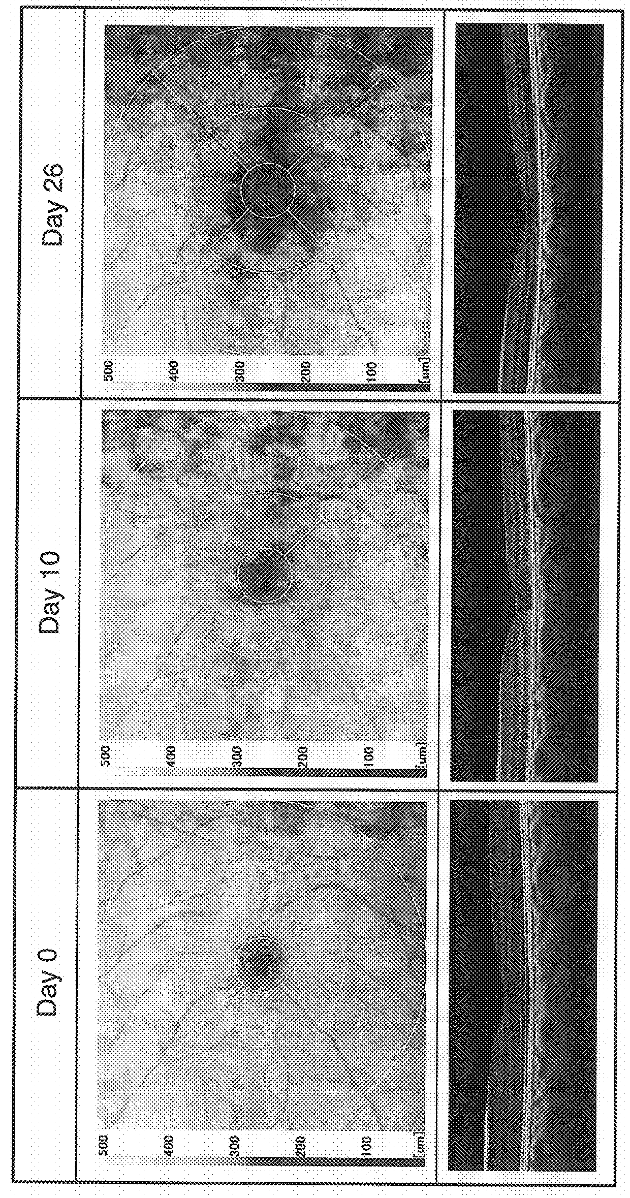
[0051]Retinal thickness of the resulting animal model intravitreally administered sodium iodate was measured prior to administration of the sodium iodate solution (day 0 of administration) and on days 10 and 26 after administration. OCT images of the retina in the vicinity of the central fossa of the macula obtained from these measurements are shown in FIG. 1. The upper row of images ...

example 2

[0053]A cynomolgus monkey model of AMD was produced to investigate whether or not the model exhibits histological characteristics unique to human AMD in the ocular fundus and central fossa of the macula.

[0054]

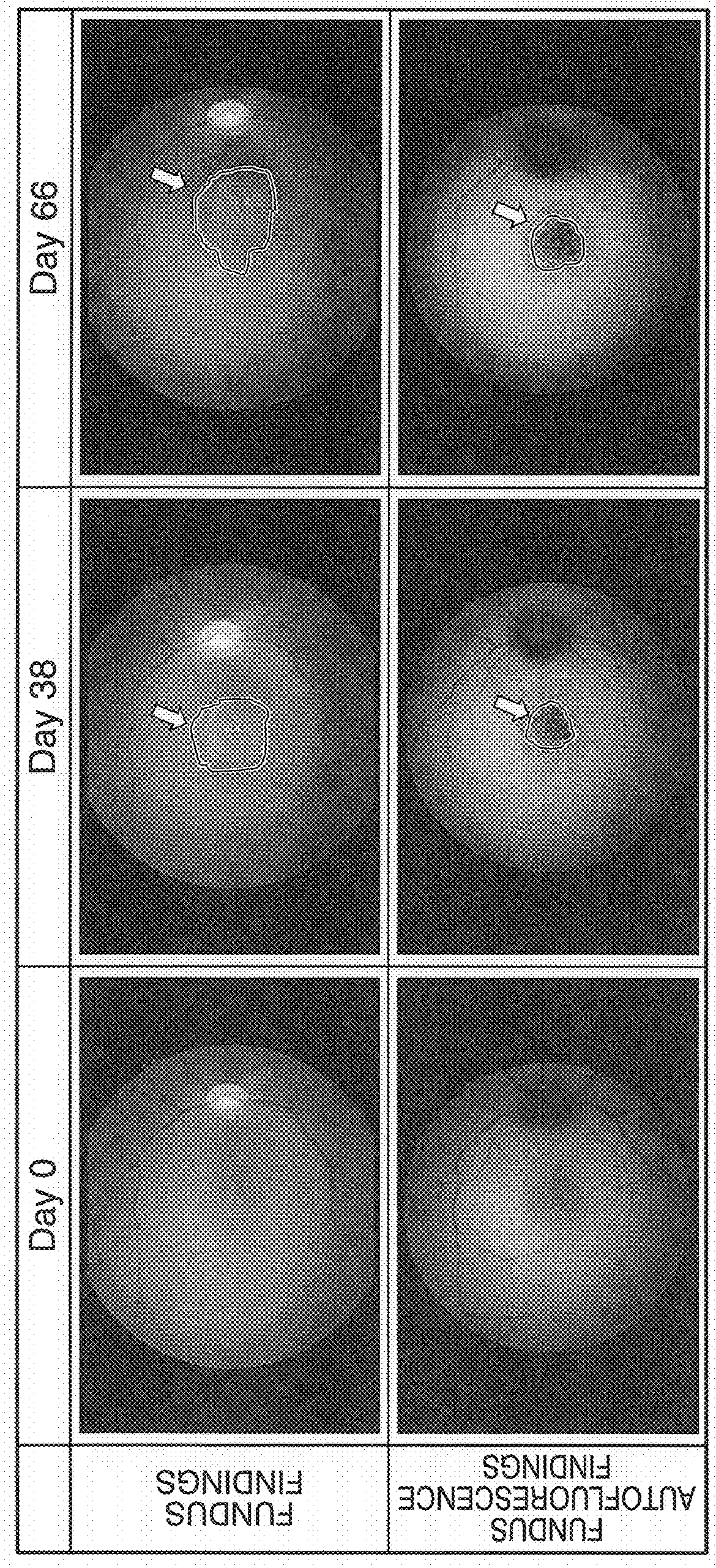
[0055]More specifically, 1.5 mg of sodium iodate per vitreous body were administered into the right eye of a 9-year-old cynomolgus monkey in the same manner as Example 1 to produce an animal model of intravitreal administration of sodium iodate followed by investigating the ocular fundus by funduscopy and fundus autofluorescence prior to administration of the sodium iodate solution (day 0 of administration) and on days 38 and 66 after administration. Funduscopy and fundus autofluorescence were carried out in accordance with routine methods using a fundus camera equipped with an autofluorescence imaging function. In addition, the animal model also underwent fluorescein angiography on day 66 after administration. Fluorescein angiography was carried out in accordance with routine ...

reference example 1

[0062]A cynomolgus monkey model of light-induced retinopathy was produced to investigate the morphology of the retinal tissue of that model by HE staining.

[0063]

[0064]One eye of a cynomolgus monkey immobilized while facing upward was illuminated for 30 minutes with blue light at an illumination intensity of 1800 lux and wavelength of 460 nm from directly overhead at a distance of 5 cm from the eye using a variable wavelength light source. This procedure was carried out for 3 days to produce an animal model of light-induced retinopathy.

[0065]

[0066]Tissue at sites surrounding the central fossa of the resulting animal model of light-induced retinopathy was subjected to HE staining to investigate the morphology of the RPE layer. HE staining was carried out in the same manner as Example 2.

[0067]Images of the HE-stained tissue at sites surrounding the central fossa are shown in FIG. 6. Significant degeneration of the RPE layer was not observed. Detachment of the photoreceptor cell layer f...


PUM


Abstract

An object of the present invention is to provide a method for producing a non-human primate model of AMD, a method for evaluating the efficacy of a test substance in the prevention or treatment of AMD using the AMD animal model produced according to this method, and a method for screening substances effective in the prevention or treatment of AMD using the aforementioned AMD animal model. The method for preparing the AMD animal model consists of administering sodium iodate into a vitreous body of a non-human primate, and the method for evaluating the efficacy of a test substance in the prevention or treatment of AMD consists of preparing a non-human primate model of AMD according to the aforementioned method for preparing an AMD animal model, and evaluating the efficacy of the test substance in the prevention or treatment of AMD using the resulting AMD animal model.

Description

[0001]The present application claims priority on the basis of Japanese Patent Application No. 2017-57515, filed on Mar. 23, 2017, and Japanese Patent Application No. 2017-242163, filed on Dec. 18, 2017, the contents of which are incorporated herein by reference in their entirety.FIELD OF THE INVENTION[0002]The present invention relates to a method for producing a non-human primate model of age-related macular degeneration (AMD), a method for evaluating the efficacy of a test substance in the prevention or treatment of AMD using an AMD animal model produced according to this method, and a method for screening substances effective in the prevention or treatment of AMD using the aforementioned AMD animal model.BACKGROUND OF THE INVENTION[0003]AMD is one of the causes of blindness, and is associated with degeneration to retinal photoreceptor cells of the macula corresponding to the center of the ocular fundus. AMD is primarily classified into an atrophic form (dry type) and exudativefor...

Claims


Application Information

Patent Timeline
IPC IPC(8): A61K49/00A01K67/027
CPCA61K49/0008A01K67/027A01K2207/20A01K2227/106A01K2267/035
Inventor TAKAMATSU, HIROYUKI
Owner HAMAMATSU PHARMA RES INC
Why Eureka
Try Eureka