
System and Method for Producing High-Resolution Magnetic Relaxation Parameter Maps

a technology of relaxation parameter and magnetic resonance imaging, which is applied in the field of system and method for producing high-resolution relaxation parameter map, can solve the problems of time-consuming and laborious process, inability to accurately identify tissue types outside of dense scars, and inability to accurately identify tissue types. the effect of low spatial resolution

Active Publication Date: 2019-01-24
NORTHWESTERN UNIV +1
View PDF0 Cites 3 Cited by
  • Summary
  • Abstract
  • Description
  • Claims
  • Application Information

AI Technical Summary

Benefits of technology

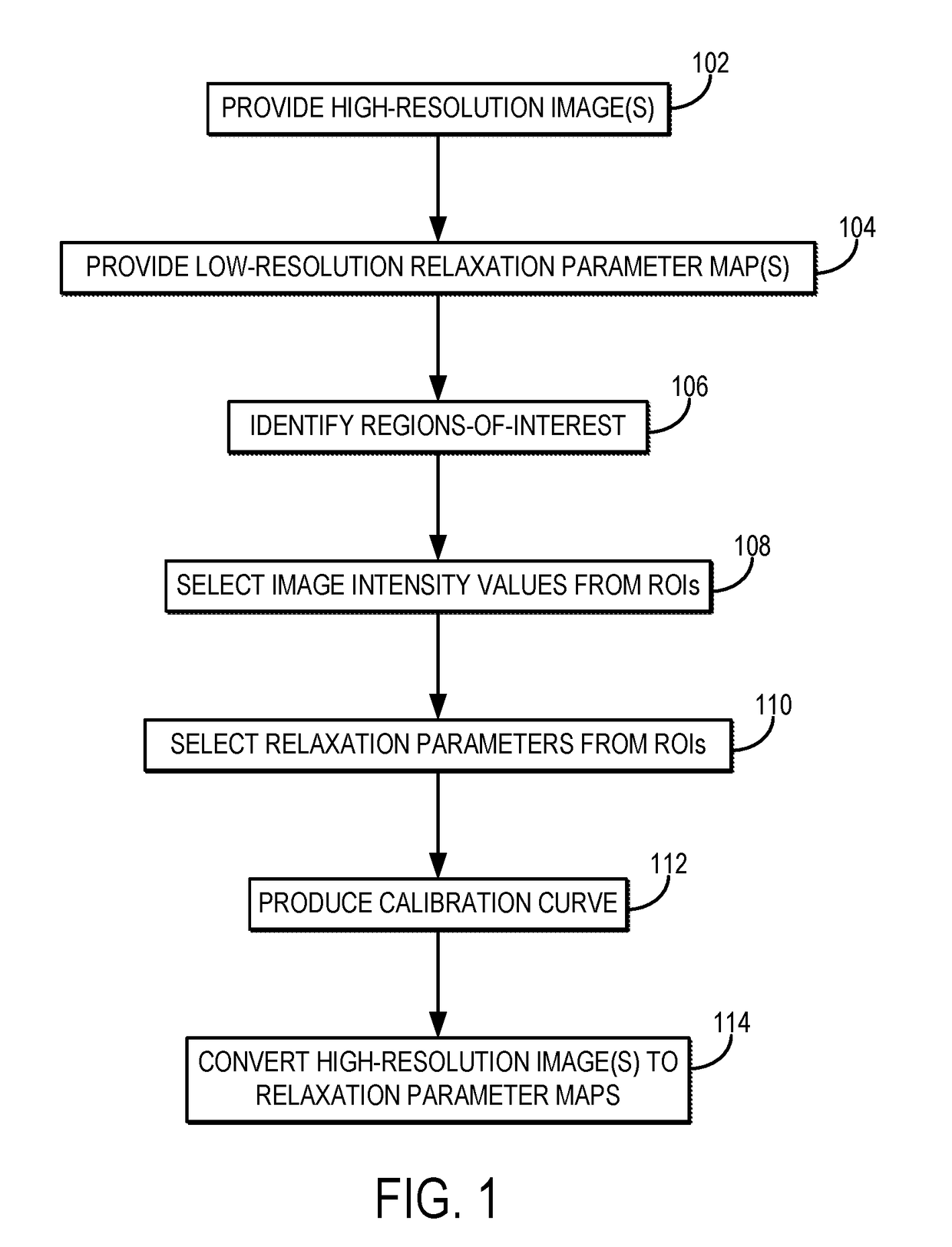
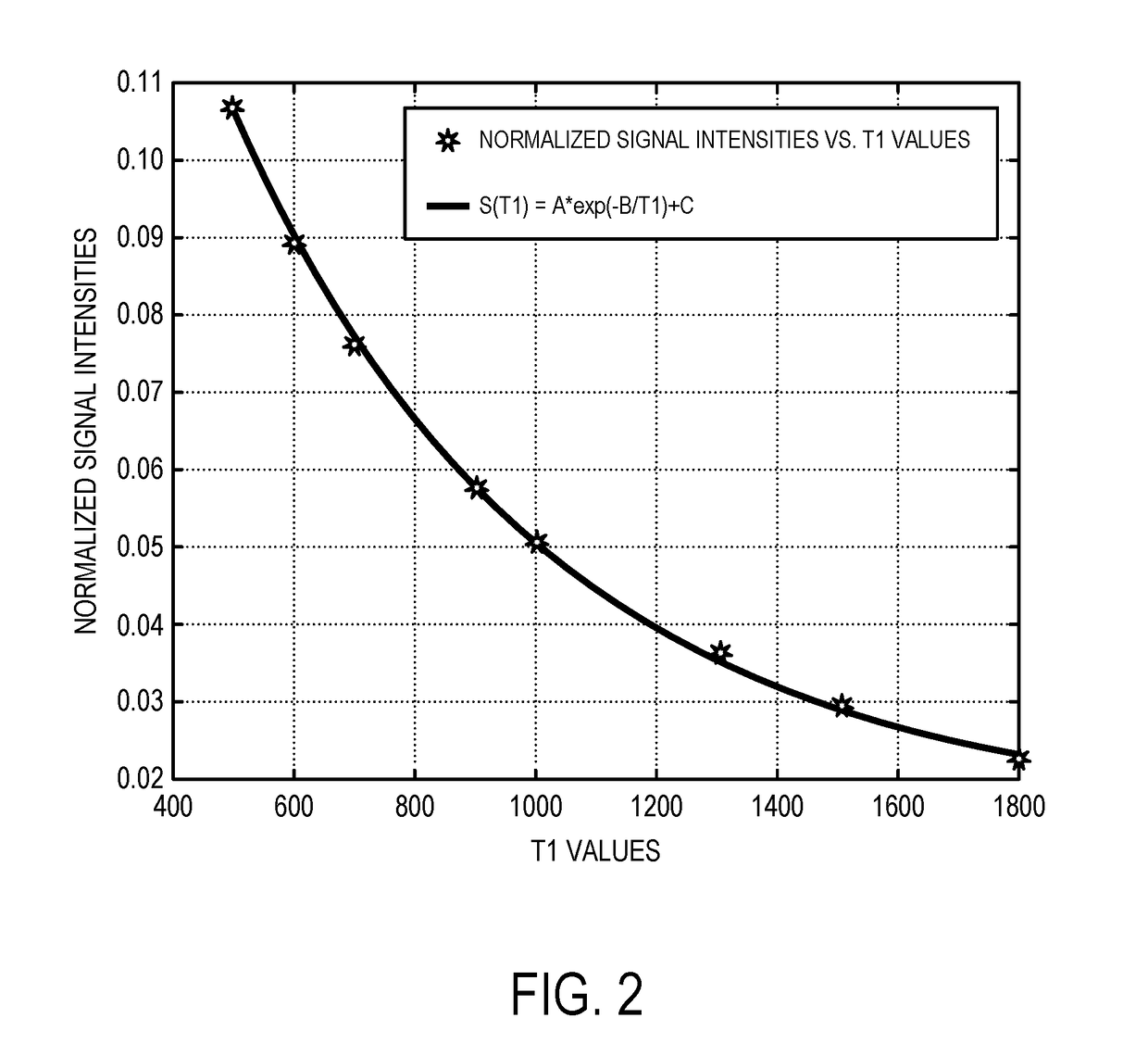
The patent describes a way to make a map of a relaxation parameter from magnetic resonance imaging (MRI) images. This is done by first getting an image of the person and a low-resolution map of the relaxation parameter. The computer system then uses these two things to create a calibration curve that connects the image to the relaxation parameter. Finally, the computer system converts the image to high-resolution using the calibration curve, resulting in a final map of the relaxation parameter. This technique creates more accurate maps of relaxation parameters, which can help in the diagnosis and treatment of various medical conditions.

Problems solved by technology

Current methods in the field to detect scar tissue and delineate areas of tissues with different characteristics are laborious and have several limitations.
Furthermore, the ability to properly identify tissue types outside of dense scar are even more difficult because thresholds for voltage collected by catheters used for scar analysis are variant between patients.
Electrical pacing from catheters in-vivo has been shown to better delineate scar tissue, but this is a time intensive process.
This technique provides information of scar tissue in relative signal intensity, which can be difficult to standardize on an individual patient basis because imaging time post contrast and heart rate are uncontrollable variables.
Due to the sampling needed at specific time points to acquire this data (in addition to the regular difficulties of cardiac imaging such as patient motion, breathing, magnetic field inhomogeneity), this can make it difficult to use the MOLLI sequence for evaluation of T1 tissue evaluation.
The acquisition of multiple sample points along the T1 recovery curve makes it difficult to achieve sub-millimeter or close to sub-millimeter isotropic resolution.

Method used


Image

  • System and Method for Producing High-Resolution Magnetic Relaxation Parameter Maps
  • System and Method for Producing High-Resolution Magnetic Relaxation Parameter Maps
  • System and Method for Producing High-Resolution Magnetic Relaxation Parameter Maps

Examples


Embodiment Construction

[0015]Described here are systems and methods for producing high-resolution three-dimensional (“3D”) relaxation parameter maps by calibrating high-resolution 3D magnetic resonance images. As one example, high-resolution longitudinal relaxation time (“T1”) maps can be generated based on images acquired using a T1-weighted pulse sequence, and as another example high-resolution transverse relaxation time (“T2”) maps can be generated based on images acquired using a T2-weighted pulse sequence. The high-resolution images can be calibrated, for example, using a lower resolution single slice relaxation parameter map. The methods described here utilize high-resolution 3D scans and low-resolution relaxation parameter maps that are commonly available on MRI systems. The calibration is a post-processing step used to create the high-resolution 3D relaxation parameter maps from these two types of scans.

[0016]The systems and methods described here include converting high-resolution 3D images to hi...


PUM


Abstract

Described here are systems and methods for producing high-resolution three-dimensional (“3D”) relaxation parameter maps by calibrating high-resolution 3D magnetic resonance images. As one example, high-resolution longitudinal relaxation time (“T1”) maps can be generated based on images acquired using a T1-weighted pulse sequence, and as another example high-resolution transverse relaxation time (“T2”) maps can be generated based on images acquired using a T2-weighted pulse sequence. The high-resolution images can be calibrated, for example, using a lower resolution single slice relaxation parameter map. The methods described here utilize high-resolution 3D scans and low-resolution relaxation parameter maps that are commonly available on MRI systems. The calibration is a post-processing step used to create the high-resolution 3D relaxation parameter maps from these two types of scans.

Description

CROSS-REFERENCE TO RELATED APPLICATIONS[0001]This application claims the benefit of U.S. Provisional Patent Application Ser. No. 62 / 276,572, filed on Jan. 8, 2016, and entitled “SYSTEM AND METHOD FOR PRODUCING HIGH-RESOLUTION MAGNETIC RESONANCE RELAXATION PARAMETER MAPS.”BACKGROUND[0002]The field of the present disclosure is systems and methods for magnetic resonance imaging (“MRI”). More particularly, the present disclosure relates to systems and methods for calibrating magnetic resonance images to produce high-resolution maps of relaxation parameters, such as longitudinal or transverse relaxation times.[0003]Cardiovascular disease is the leading global cause of death, accounting for 17.3 million deaths per year. Fibrosis is an important marker in many types of heart disease, including ischemic heart disease, atrial fibrillation, cardiac amyloidosis, cardiomyopathy, myocarditis, and aortic stenosis. Therefore, the ability to characterize and quantify the three-dimensional architect...

Claims


Application Information

Patent Timeline
Patent Type & Authority Applications(United States)
IPC IPC(8): G01R33/56G01R33/44G01R33/50G01R33/58G01R33/483
CPCG01R33/5602G01R33/443G01R33/50G01R33/58G01R33/483
Inventor NG, JASONPARIKH, PARASCARROLL, TIMOTHY J.LEE, DANIEL C.
Owner NORTHWESTERN UNIV
Learn More