Devices and methods for simulating a function of a liver tissue
a technology of liver tissue and microfluidics, which is applied in the field of microfluidic devices, can solve the problems of limited in vitro hepatic model systems, limited long-term culture systems, and limitations of conventional hepatic model systems, and achieve the effect of increasing the density of ecm fibers and reducing the deposition of ecm materials
- Summary
- Abstract
- Description
- Claims
- Application Information
AI Technical Summary
Benefits of technology
Problems solved by technology
Method used
Image

Examples
example 1
er-On-a-Chip Device Design 1 and Characterization
[0343]Hepatocytes, unlike other cell types, do not simply adhere to a surface and proliferate upon thawing. To identify suitable cell sources, cryopreserved vials were evaluated for their adhesion ability, their ability to retain their in vivo like morphology, and / or their ability to form bile canaliculi network (for biliary clearance determination). Typically, in 2D static culture hepatocytes flatten out and lose the cobblestone morphology.
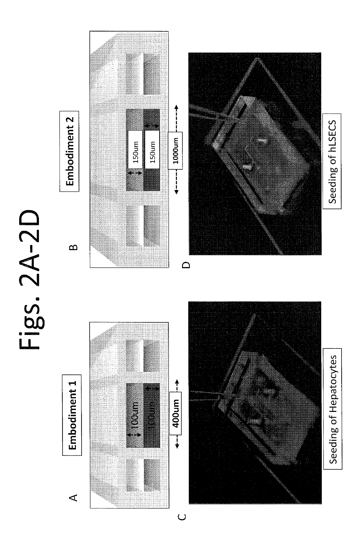
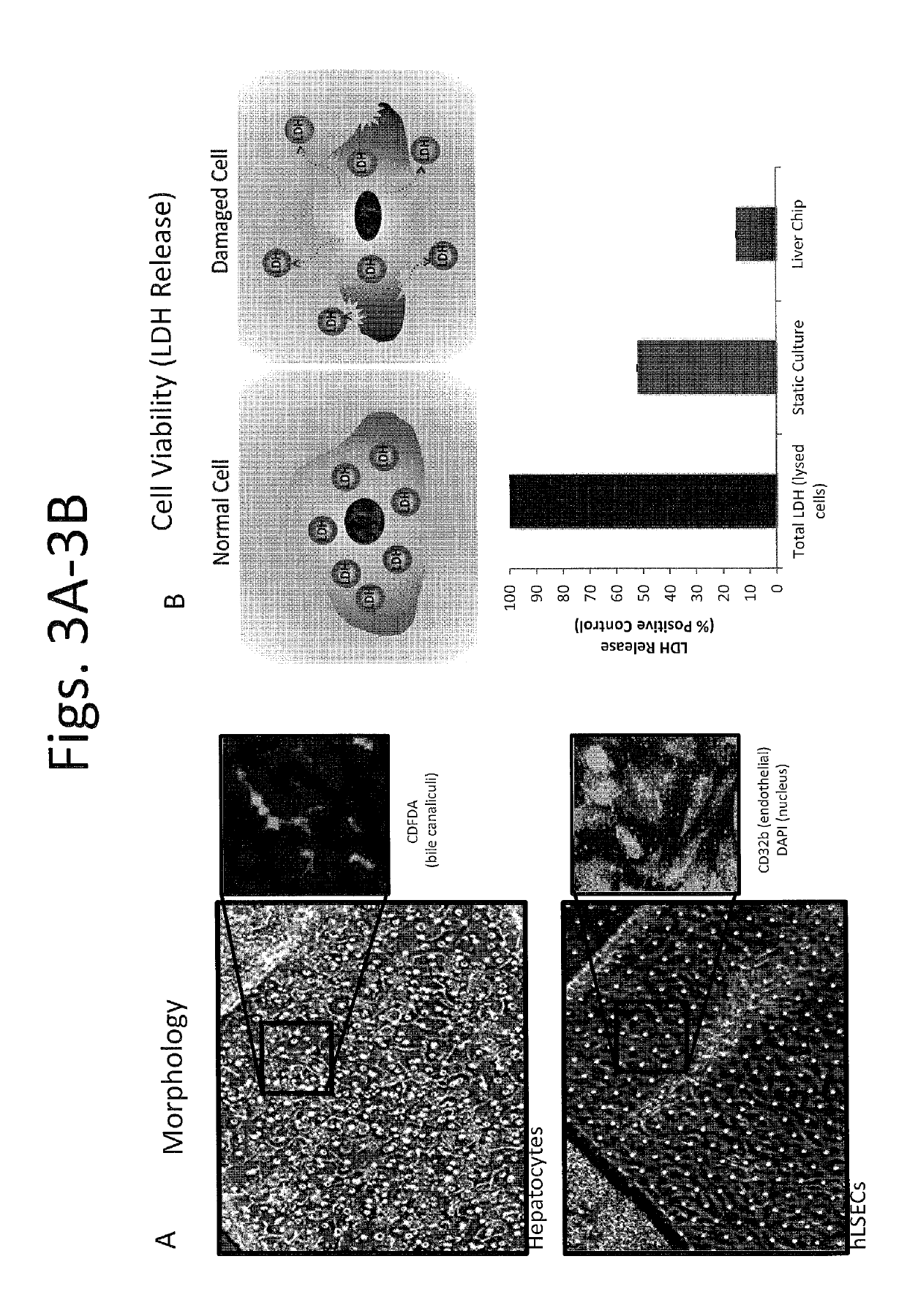
[0344]After identifying suitable cell sources of hepatocytes, the hepatocytes were cultured on a first side of the membrane (coated with collagen I at approx. 100 ug / mL) facing a first chamber of the device, e.g., as shown in FIG. 2B or FIG. 7B, to permit growth of a confluent monolayer within the first chamber. FIG. 3A is a set of images showing hepatocytes (top panel) and hLSECs (bottom panel) from Day 13. It is noted that the cells were able to continue to grow for at least about 3 weeks or long...
example 2
er-On-a-Chip Device Design 2 and Characterization
[0352]As hepatocytes are much more cuboidal than epithelial cells of other tissue, e.g., alveolar flat cell type, the height of the chamber in which the hepatocytes are cultured can be increased to accommodate the cell size as well as the width so that much more confluent monolayers of hepatocytes can be achieved. For example, in one embodiment, the hepatocytes can be cultured in the top chamber of the device shown in FIG. 78. As described in Example 1, CYP3A4 activity of the hepatocytes growing in the device of FIG. 7B (referred to as “Liver chip v2”) was measured over a period of time, and compared to the cells grown in the device of FIG. 7A (referred to as “Liver chip v1”). FIG. 8 shows that CYP3A4 activity levels steadily increased in Liver chip v1, while the Liver chip v2 showed comparable activity levels to fresh hepatocytes and the CYP3A4 activity was maintained in Liver chip v2 for at least about 2 weeks. Similarly, hepatocyte...
example 3
-On-a-Chip Device Design and Characterization
[0356]Similar to development of human liver-on-a-chip devices as described in Examples 1-2, suitable cell sources of rat hepatocytes and rat liver sinusoidal endothelial cells (rLSECs) were first identified. Cryopreserved vials were evaluated for their adhesion ability, their ability to retain their in vivo like morphology, and / or their ability to form bile canaliculi network (for biliary clearance determination). Rat hepatocytes from Xenotech Lot: XT1310140 and Biopredic Lot: HEP139031-T and rLSECs from Cell Biologics Lot: R1072 were selected to develop rat liver-on-a-chip devices. See FIGS. 23A-23B for characterization of hepatic function of rat hepatocytes obtained from XenoTech and Biopredic.
[0357]In order to achieve long-term maintenance (e.g., viability and function) of a co-culture model of rat hepatocytes and rLSECs in a device as shown in FIG. 7B, e.g., for at least two weeks or longer, the inventors have identified four optimiza...
PUM
 Abstract
Description
Claims
Application Information
 - R&D
- Intellectual Property
- Life Sciences
- Materials
- Tech Scout
- Unparalleled Data Quality
- Higher Quality Content
- 60% Fewer Hallucinations
Browse by: Latest US Patents, China's latest patents, Technical Efficacy Thesaurus, Application Domain, Technology Topic, Popular Technical Reports.
© 2025 PatSnap. All rights reserved.Legal|Privacy policy|Modern Slavery Act Transparency Statement|Sitemap|About US| Contact US: help@patsnap.com