Method for measuring a viscoelastic parameter of a human or animal organ

a viscoelastic parameter and human or animal organ technology, applied in the field of biological tissue viscoelastic parameter measurement, can solve the problems of low signal-to-noise ratio, other infections of the liver are currently more difficult to assess, and known technical solutions provide measurements with low signal-to-noise ratios, etc., to achieve the effect of increasing the measured signal, and increasing the signal-to-noise ratio

Active Publication Date: 2019-08-29
ECHOSENS SA
View PDF0 Cites 1 Cited by
  • Summary
  • Abstract
  • Description
  • Claims
  • Application Information

AI Technical Summary

Benefits of technology

[0031]One advantage of the invention is that it increases the measured signal, since several pulses at different frequencies are sent into the tissue. Measurements with a higher signal-to-noise ratio are then made. In addition, this increase of the signal-to-noise ratio is accomplished without increasing the intensity associated with each pulse: the total transmitted power is shared by several pulses spread over a wide range of frequencies. The range of accessible frequencies is the bandwidth of the ultrasound transducer.
[0032]In the case of the human liver, the reference frequency to measure steatosis is 3.5 MHz (central frequency). In a range of several MHz around this frequency, coefficient α(f) varies approximately linearly with frequency f.
[0033]By virtue of the invention it is possible to measure α(f) with a better signal-to-noise ratio for several values of f around the central frequency of 3.5 MHz and, using for example a simple linear regression, to obtain a more precise estimate of reference value α(f=3.5 MHz).
[0040]Fc is the central frequency of the ultrasound transducer, P is equal to 5 and the chosen frequencies are 0.5*Fc<F1<0.7*Fc; 0.7*Fc<F2<0.9*Fc; 0.9*Fc<F3<1.1*Fc; 1.1*Fc<F4<1.3*Fc; 1.3*Fc<F5<1.5*Fc;
[0044]means of recording and processing the reflected ultrasound signals able to determine at least one viscoelastic parameter of the organ.
[0047]measurement of a parameter of attenuation of the ultrasound signals with the method according to the invention described in the previous paragraphs.

Problems solved by technology

Other infections of the liver are currently more difficult to assess.
However, the known technical solutions provide measurements with a signal-to-noise ratio which can be very low, particularly in the case of obese patients—whose livers often have steatosis—due to the high attenuation of the ultrasound pulses in the subcutaneous tissues (in particular fat), and in the medium to be measured itself.

Method used


Image

  • Method for measuring a viscoelastic parameter of a human or animal organ
  • Method for measuring a viscoelastic parameter of a human or animal organ
  • Method for measuring a viscoelastic parameter of a human or animal organ

Examples


Embodiment Construction

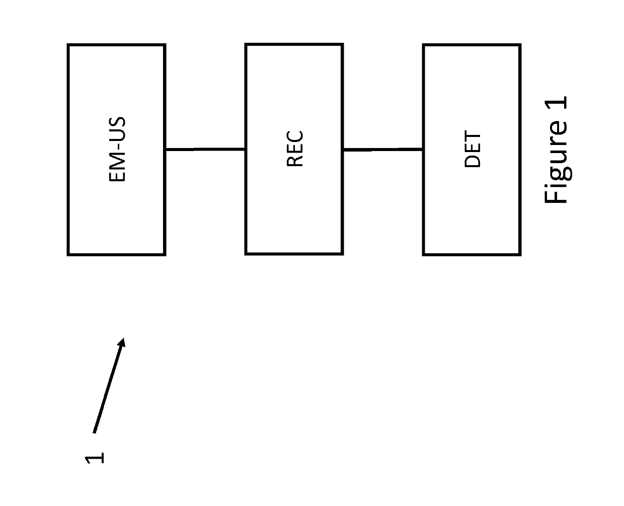
[0061]FIG. 1 illustrates the different steps of first method 1 of the invention. Method 1 in this case includes three steps:[0062]an emission EM-US by an ultrasound transducer US of a plurality of ultrasound shots, which are propagated in the organ to be characterised,[0063]a recording REC by the same ultrasound transducer US of the reflected ultrasound signals,[0064]a determination DET of at least one viscoelastic parameter of the organ from the recorded signals.

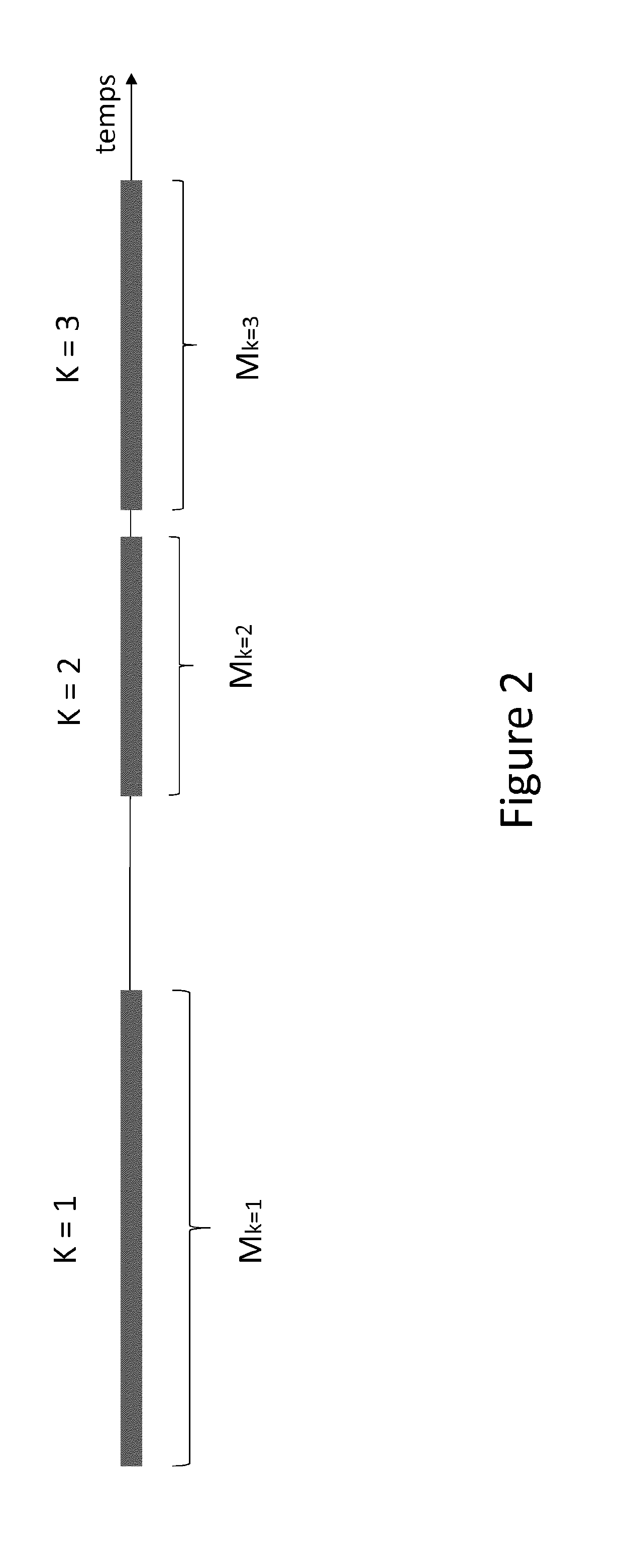
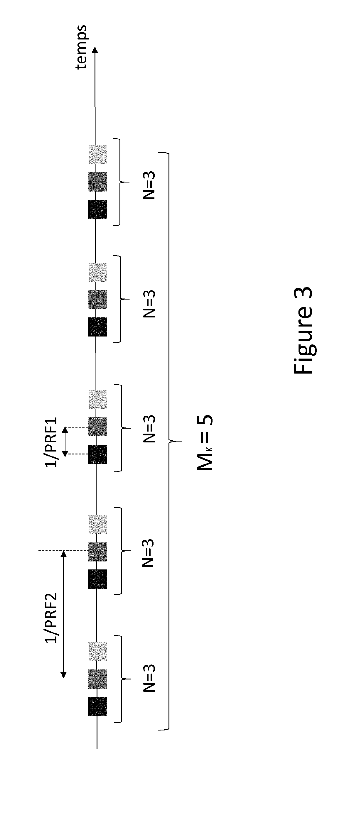
[0065]Advantageously, this implementation enables a parameter such as the attenuation of the ultrasound signals to be measured. This parameter is particularly significant in the case of the human liver since it is related to the presence of steatosis. The structure and characteristics of the plurality of ultrasound shots are represented in FIGS. 2 and 3.

[0066]FIG. 2 shows that the plurality of shots is formed by K groups of ultrasound shots. For example, in the case shown in FIG. 2, there are K=3 groups of ultrasound shots....


PUM


Abstract

A method for measuring a viscoelastic parameter of an organ, includes emitting ultrasonic shots by an ultrasonic transducer; receiving by the transducer and recording the reflected ultrasonic signals; determining a viscoelastic parameter of the organ based on the recorded ultrasonic signals. The ultrasonic shots are formed by K groups of shots, separated temporally, K being greater than or equal to 1. Each K group is formed by the repetition, with a rate of PRF2, of MK blocks of ultrasonic shots, MK being greater than or equal to 1; each MK block is composed of N ultrasonic shots, N being greater than or equal to 1, PRF1 being the rate of emission of the N shots when N is chosen greater than 1; the N ultrasonic shots are distributed over P frequencies, P being between 1 and N, at least two ultrasonic shots belonging to two different blocks having different frequencies.

Description

FIELD OF THE INVENTION[0001]The present invention concerns the field of measurement of the viscoelastic properties of a biological tissue. More specifically, the invention relates to a method for measuring a viscoelastic parameter using multi-frequency ultrasound pulses.STATE OF THE ART[0002]Several non-invasive techniques for measuring the viscoelastic properties of human or animal organs have been developed.[0003]Of these techniques, pulse elastography is particularly effective in monitoring infections in soft tissues, and particularly the human liver. For example, the applicant has developed and marketed a device, Fibroscan™, which is now a reference for diagnosing and monitoring hepatic fibrosis. This device is illustrated, in particular, in patents EP 1 169 636 and EP 1 531 733 filed by the applicant. This infection can be the consequence of illnesses such as hepatitis or cancer of the liver, and it is characterised by a loss of elasticity of the liver. The technology developed...

Claims


Application Information

Patent Timeline
Patent Type & Authority Applications(United States)
IPC IPC(8): A61B8/08A61B8/00G01S7/52
CPCA61B8/485A61B8/085A61B8/54G01S7/52042G01S7/52022A61B8/5207A61B8/5223A61B8/08A61B8/44A61B8/4483G01S7/52036
Inventor AUDIERE, STÉPHANESANDRIN, LAURENT
Owner ECHOSENS SA
Why Eureka
Try Eureka