
Device for utilizing transmission ultrasonography to enable ultrasound-guided placement of central venous catheters

a technology of transmission ultrasonography and catheters, applied in the field of medical imaging, can solve the problems of unsatisfactory radiation involved in this procedur

Active Publication Date: 2019-12-19
THE JOHN HOPKINS UNIV SCHOOL OF MEDICINE
View PDF0 Cites 0 Cited by
  • Summary
  • Abstract
  • Description
  • Claims
  • Application Information

AI Technical Summary

Benefits of technology

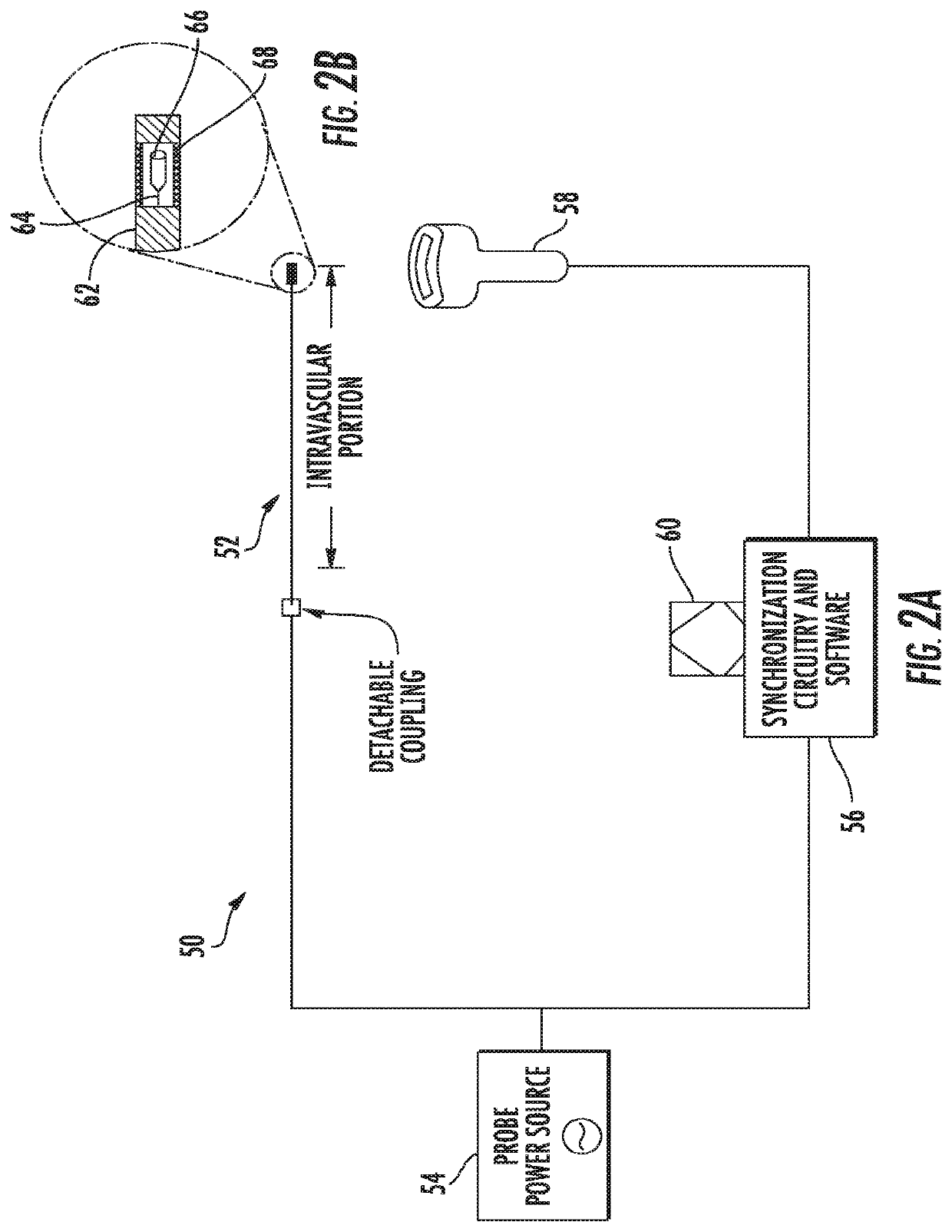
The present invention provides a system for ultrasonography in blood vessels using an intravascular probe and a detection probe. This system can be used to diagnose and treat blockages in blood vessels. The intravascular probe produces ultrasonic energy and the detection probe detects the energy and sends a signal to a computing device for processing and display. The computing device can display information about the blood vessel being examined. The intravascular probe and the catheter can be piezoelectric devices. The detection probe can be a blanket array or a single probe on a robotic arm. The system can help improve the diagnosis and treatment of blockages in blood vessels.

Problems solved by technology

However, the amount of radiation involved in this procedure is not ideal, particularly for small children.

Method used


Image

  • Device for utilizing transmission ultrasonography to enable ultrasound-guided placement of central venous catheters
  • Device for utilizing transmission ultrasonography to enable ultrasound-guided placement of central venous catheters
  • Device for utilizing transmission ultrasonography to enable ultrasound-guided placement of central venous catheters

Examples


example

[0032]The follow exemplary implementation of an ultrasound probe according to an embodiment of the present invention is included merely as an example, and is not considered to be limiting. Any other implementation known to or conceivable by one of skill in the art could also be used.

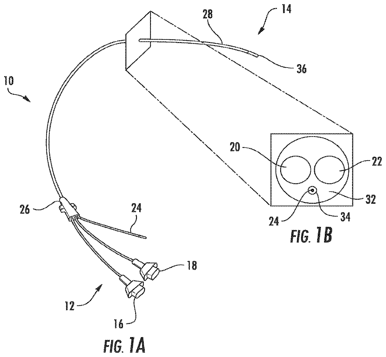
[0033]An intravascular transmission ultrasonography probe was custom fabricated, in which a piezoelectric transducer actively emits signal that can be detected by a standard handheld probe, rather than relying solely on reflected acoustic waves to create an image. A miniaturized element was integrated into a flexible catheter that was introduced by vascular sheath into the central veins of an immature pig, as illustrated in FIG. 7A.

[0034]Catheter tip location was noted to blink on B-mode ultrasound with a standard handheld ultrasound probe, as illustrated in FIG. 7B. Tip location along the vessel axis was estimated using the probe freehand and marked with radiopaque beads within 1 cm of its superior / infe...


PUM


Abstract

Ultrasound is typically performed with the use of an external transcutaneous probe that emits ultrasonic energy and measures the timing of reflected waves, thus allowing measurement of fluid flow or formation of a two-dimensional image. Clinically, the modality is limited by attenuation of the signal with increasing depth of penetration of the ultrasound into tissues, as well as by similarities in echo-genicity between different tissues. The present invention includes a system in which an intravascular probe actively transmits ultrasound that can then be detected by in-line piezoelectric transducers without reflection. This signal can be overlaid onto traditional B-mode or Doppler representation. Furthermore, the present invention includes a signal processing system that will display a real-time graphical representation of the vascular anatomy in order to assist the surgeon or procedural radiologist in the placement of central venous catheters.

Description

CROSS REFERENCE TO RELATED APPLICATION[0001]This application claims the benefit of U.S. Provisional Patent Application No. 61 / 872,903 filed on Sep. 3, 2013, which is incorporated by reference, herein, in its entirety.FIELD OF THE INVENTION[0002]The present invention relates generally to medical imaging. More particularly, the present invention relates to a device for image guided placement of a central venous catheter.BACKGROUND OF THE INVENTION[0003]Central venous catheters have many uses and are used in patients of all ages, including pediatric patients. Central venous catheters are generally inserted using the Seldinger technique. First, a blunt guidewire is passed through a needle inserted into the vein. The progress and position of the guidewire can be monitored using fluoroscopy. After the guidewire is positioned, a stiff introducer is inserted, the guidewire is removed, and the catheter is passed into the vein within the introducer. Once the catheter is positioned, more fluor...

Claims


Application Information

Patent Timeline
Patent Type & Authority Applications(United States)
IPC IPC(8): A61B8/12A61B8/08A61B8/00
CPCA61B8/445A61B8/0841A61B8/12A61B8/4483A61B8/4218
Inventor ABDULLAH, FIZANGOLDSTEIN, SETHBOCTOR, EMAD
Owner THE JOHN HOPKINS UNIV SCHOOL OF MEDICINE
Learn More