Operating a medical image recording device

a medical image and recording device technology, applied in image data processing, biological neural network models, radiation therapy, etc., can solve the problems of measurement protocols that do not solve problems, complex systems of modern medical image recording devices, and difficult operation, so as to achieve the effect of accurately describing patients

Active Publication Date: 2019-12-19
SIEMENS HEALTHCARE GMBH
View PDF0 Cites 1 Cited by
  • Summary
  • Abstract
  • Description
  • Claims
  • Application Information

AI Technical Summary

Benefits of technology

[0022]Specifically, the registration may be effected using sensor data from at least one sensor directed at the patient and/or image data of the patient recorded by the image recording device and/or based on patient data retrieved from an information system and/or based on patient location data obtained from an embodiment of a patient couch of the image recording device. In a specific embodiment, a camera (e.g., a 3D camera and/or an infrared camera and/or terahertz camera, and/or an ultrasound sensor) may be used as a sensor with other embodiments also being possible. Specifically, a multiplicity of methods may therefore be used to effect a registration of the patient with a coordinates system of the image recording device. The registration is, for example, manifested specifically in a registration of the patient model that is adapted to the current patient with this coordinates system.
[0023]Initially, a camera-based optical registration, where the camera replicates the patient on the patient couch and generates a correlation of the patient outline relative to the patient couch, which, for example, also describes the location of the patient (e.g., “head first”, “feet first”, “supine”, “prone”, etc.), may be provided. The use of an infrared camera has the advantage of being able to see through any patient covering in a sterile manner. Ultrasound reco

Problems solved by technology

Modern medical image recording devices are complex systems for which operation represents a considerable challenge to users required to obtain suitable or even optimal image quality during an i

Method used


Image

  • Operating a medical image recording device
  • Operating a medical image recording device

Examples


Embodiment Construction

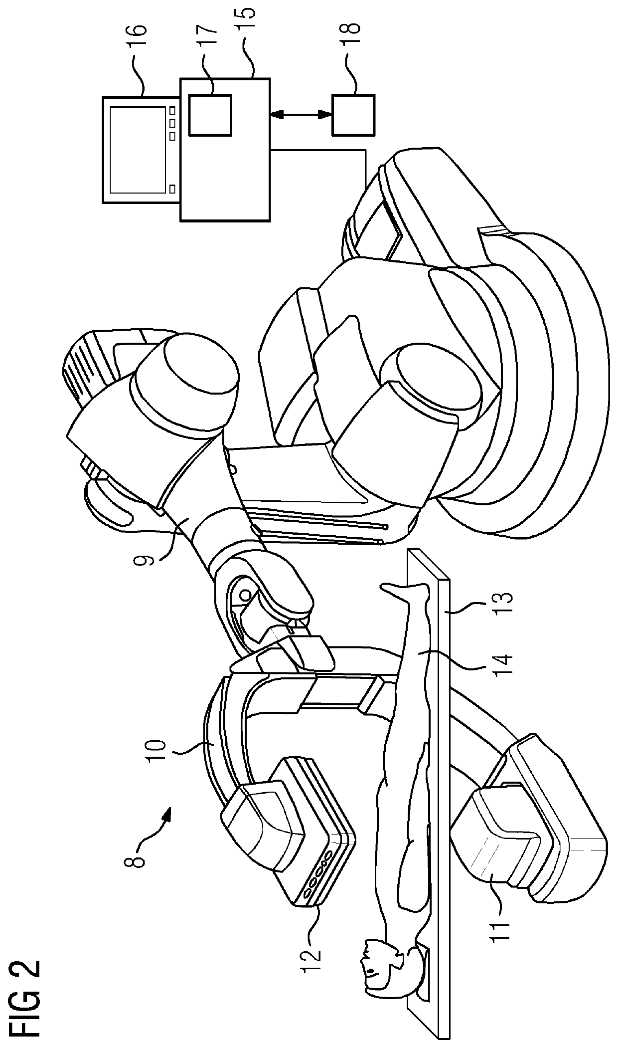
[0045]Exemplary embodiments are discussed in the following with reference to an X-ray device as an image recording device. Application to other image recording devices of greater complexity (e.g., magnetic resonance devices) may also be provided.

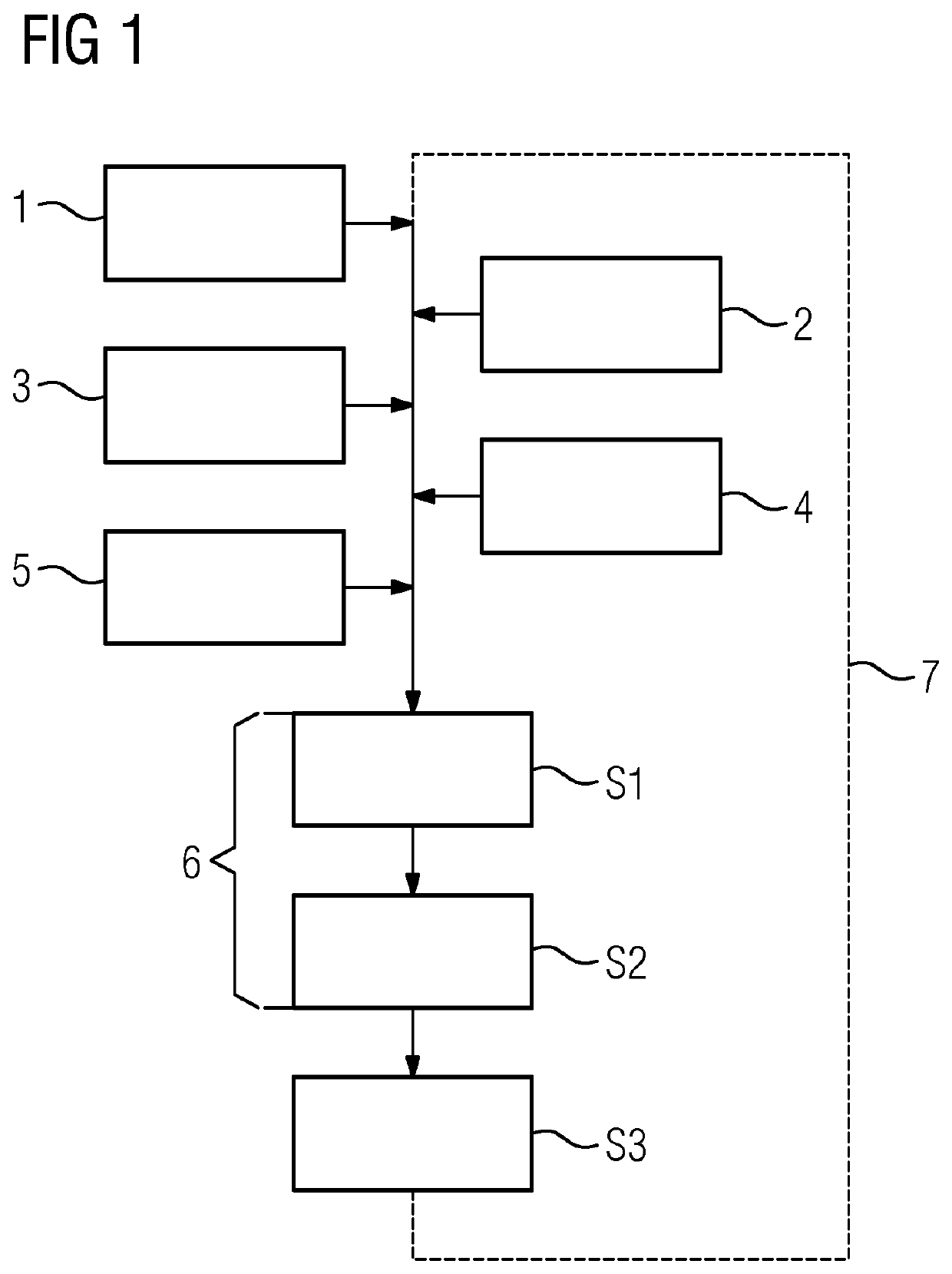
[0046]Optimal physical and image processing operating parameters are to be selected in a fully automatic manner by a control device of the image recording device based on some or all of the available input information, such as, for example: a patient model adapted to the current patient; registration of the patient model and therefore the patient with a coordinates system of the image recording device; knowledge of the image recording purpose; information relating to a current examination region (e.g., organ of interest (OOI)); information relating to implants, direct radiation, location of filters / collimators, inclusion of expected movements in the examination region; and knowledge relating to medical instruments used or the medical instrum...


PUM


Abstract

Operating a medical image recording device in the context of an image recording routine for a patient is provided. The image recording routine serves an image recording purpose. The image recording device is controlled based on operating parameters implemented by a controller of the image recording device. The operating parameters are identified completely automatically by an identification algorithm from input data describing at least the patient in the form of a patient model and/or the image recording purpose, and from a registration of the patient with a coordinates system of the image recording device. The operating parameters are used to control the image recording device. An examination region that is to be recorded for the patient and the image recording purpose are derived based on the registration.

Description

[0001]This application claims the benefit of EP 18178021.4, filed on Jun. 15, 2018, which is hereby incorporated by reference in its entirety.BACKGROUND[0002]The present embodiments relate to operating a medical image recording device in the context of an image recording routine for a patient.[0003]Modern medical image recording devices are complex systems for which operation represents a considerable challenge to users required to obtain suitable or even optimal image quality during an image recording routine for a patient. It is therefore customary in the prior art for users to be offered a choice of manually selectable predefined measurement protocols, so that users may select an appropriate measurement protocol based on the image recording purpose of the current image recording routine. Such measurement protocols are also referred to as “organ programs” (e.g., when X-ray devices are used as image recording devices).[0004]The measurement protocols, which define operating paramete...

Claims


Application Information

Patent Timeline
IPC IPC(8): A61B90/00A61B6/03A61B6/00A61N5/10A61B34/20G06T7/32
CPCA61B90/36G16H30/40G06N3/08A61N2005/1059A61B2034/2055G16H50/50A61B6/5217G06T7/38A61B2090/364A61B6/032A61B6/547G06T7/32A61B34/20A61B6/5229A61N5/1049A61B6/467A61B6/5294A61B6/545G16H40/63
Inventor SPAHN, MARTINBERNHARDT, PHILIPPBÖHM, STEFANSTOWASSER, BORISOBLER, RICHARDTASHENOV, STANISLAVLEIBLEIN, RUDOLFBERTING, ANDREASLENDL, MARKUSBARUTH, OLIVER
Owner SIEMENS HEALTHCARE GMBH
Why Eureka
Try Eureka