
Endoscope tip attachment device

a technology of endoscope and attachment device, which is applied in the field of endoscope accessories, can solve the problems of inability to allow a clinician to accurately visualize, inability to safely and inability to effectively so as to reduce the time taken by the clinician, the effect of increasing the resistance to force upon withdrawal and being more complian

Active Publication Date: 2020-02-06
HOYA CORP
View PDF0 Cites 0 Cited by
  • Summary
  • Abstract
  • Description
  • Claims
  • Application Information

AI Technical Summary

Benefits of technology

[0016]Accordingly, an improved endoscope attachment device is needed that is more compliant upon insertion and has a higher resistance to force upon withdrawal. Such a device may be capable of safely and effectively reducing the time taken for the clinician to perform an endoscopic procedure and for increasing the effectiveness of the procedure.
[0017]The device of the present disclosure aims to overcome the limitations of the prior art by facilitating one or more of the following: Low resistance in an insertion direction; more effective opening of folds on withdrawal, steadying and / or centering the endoscope tip's position during a medical procedure; reducing the potential for mucosal trauma; and / or providing better physical and / or visual access around colonic folds.Solution to Problem

Problems solved by technology

While colonoscopic and enteroscopic examinations may be effective techniques to assess the state of health of an internal body region, they may cause complications and, in some instances may fail to allow a clinician to accurately visualize a region of interest.
For example, the clinician may not be able to complete a procedure may fail to detect a polyp, lesion, or other tissue, or may cause injury to a body lumen in which the endoscope is inserted, e.g., via the application of a traumatic force, which may result in inflammation, burns, bleeding, scarring, perforation, or other injury.
Endoscopic procedures may be time consuming for patients and medical personnel alike, depending upon how difficult it is to advance a scope through the body lumen or to view the surrounding region of the body lumen.
Increased procedure time requires a patient to be sedated for longer periods may increase patient discomfort, and thus may increase recovery time.
Increased procedure time further cuts down on the number of procedures that a given team of clinicians can perform in one day and limits the use of an operating room.
Anatomical and technological limitations may also contribute to the difficulties of these procedures.
First, the anatomy of a body lumen, e.g., the colon may be tortuous, and the lining thereof may be uneven.
As the tip of the endoscope passes along the lumen of the colon, these folds may hamper the clinician's ability to visualize the entire surface of the mucosa and, in particular, to detect pre-malignant and malignant lesions and polyps located along these folds.
Second, the tip position of the endoscope may be difficult to maintain once a lesion or polyp is detected and / or during a therapeutic, diagnostic, or biopsy procedure.
This movement and / or the effect of gravity may cause the clinician to become disoriented, lose visualization, or lose positioning.
Additionally, the tortuous nature of the gastrointestinal tract may make it difficult for the clinician to navigate the endoscope to the region of interest.
The turns of the bowel, folded surface of the colon, and effects of gravity may cause the endoscope to bump and press on the body lumen as the endoscope is advanced or withdrawn.
This may lead to stretching of the bowel, perforation, bleeding, trauma to the mucosa, inflammation, or other injury.
As a result, the patient may experience pain, the patient's recovery time may increase, procedure time may increase, or the procedure may even need to be aborted prematurely.
However, these devices may be complex to manufacture and use due to the need for inflation and deflation mechanisms and the delicateness of the expanding portions.
Additionally, active balloons that form a permanent part of an endoscope make scope-reprocessing (e.g., high level cleaning and disinfection) more difficult.
However, the protrusions of those devices typically provide very similar stiffness and resistance to force in the direction of insertion and the direction of withdrawal.
If the protrusions are stiff upon insertion, this may cause increased insertion resistance, which then might cause the scope to loop and stretch the colon walls.
This might produce mucosal trauma as the endoscope is inserted or withdrawn.
Additionally, force applied by the tips of the protrusions to discrete surface areas of the wall of the body lumen may increase mucosal trauma or cause perforation.
On the other hand, if the protrusions are not stiff on withdrawal, they may not be capable of opening the colonic folds and may not help with visualization of the regions adjacent to the folds, as intended.

Method used


Image

  • Endoscope tip attachment device
  • Endoscope tip attachment device
  • Endoscope tip attachment device

Examples


Embodiment Construction

[0066]Reference will now be made in detail to the exemplary embodiments of the present disclosure described below and illustrated in the accompanying drawings. Wherever possible, the same reference numbers will be used throughout the drawings to refer to same or like parts.

[0067]For purposes of this disclosure, an “endoscope” may refer to any suitable type of scope for insertion into a patient during a medical procedure. Endoscopes may include, for example, colonoscopes, duodenoscopes, gastroscopes, sigmoidoscopes, enteroscopes, ureteroscopes, and bronchoscopes. The term “procedure” broadly refers to the insertion of an endoscope into a patient for any purpose, including, but not limited to, surgery, biopsy, diagnosis, treatment, visualization, implantation or removal of a device, suction, or insufflation.

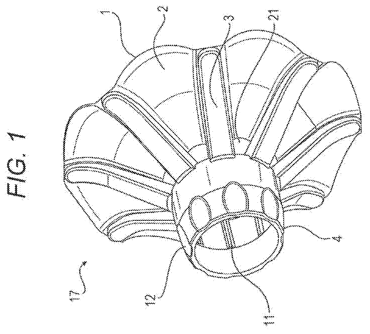
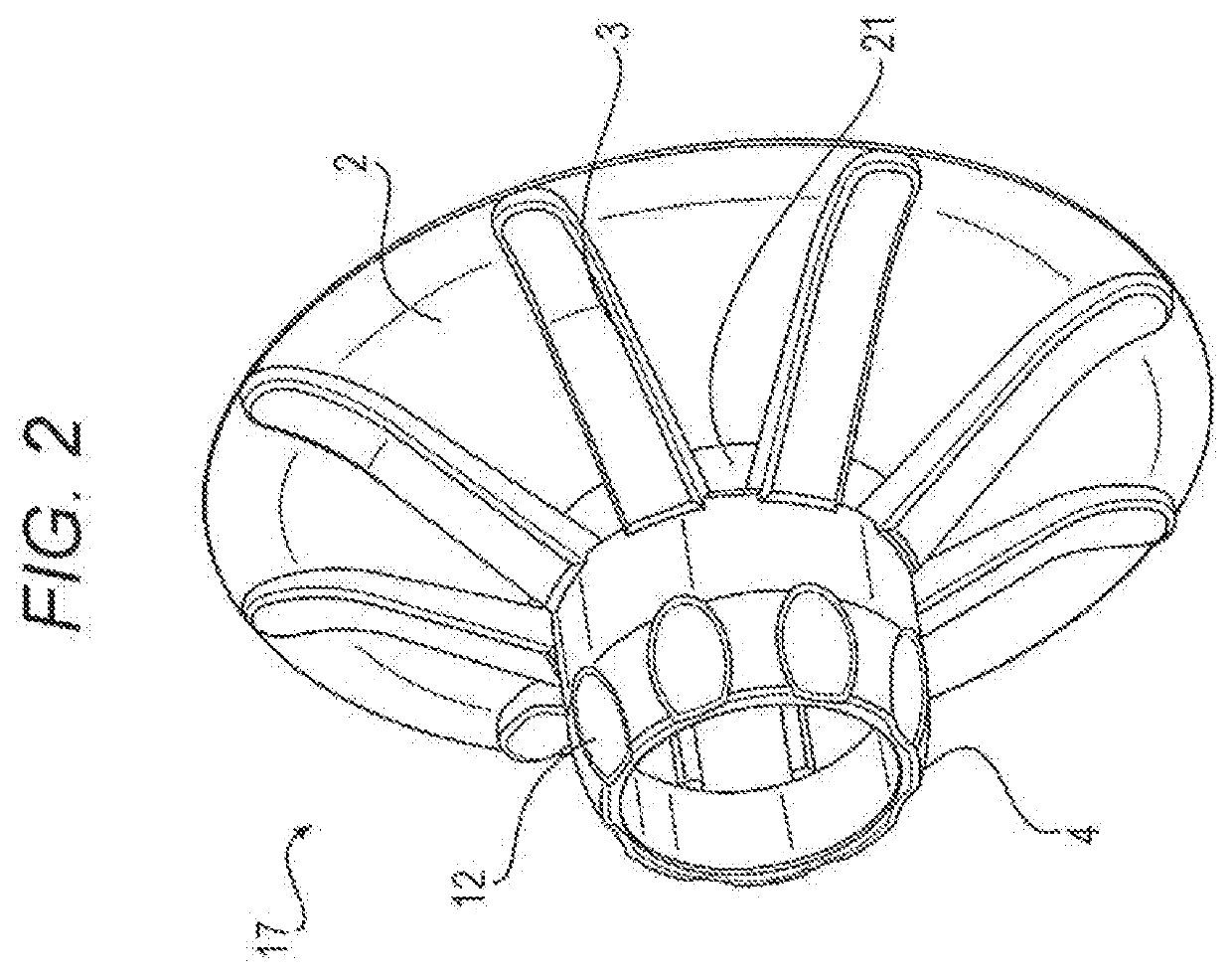
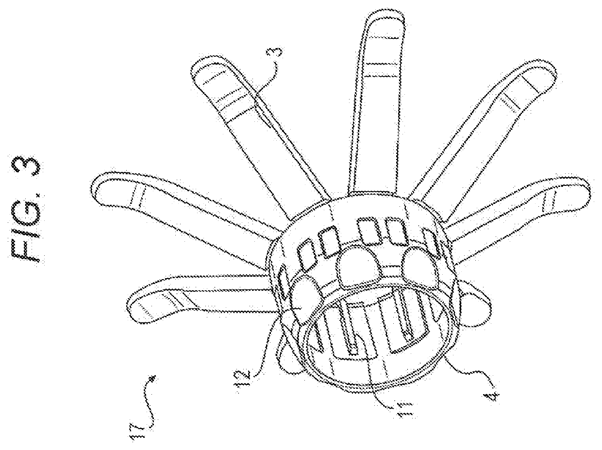
[0068]Prior to providing a detailed description, the following overview generally describes the contemplated embodiments. Endoscope tip assembly 17 of the current disclosure is con...


PUM


Abstract

Embodiments of the disclosure include an endoscope tip assembly for use on an endoscope, during procedures. The endoscope tip assembly may include a base configured to receive an endoscope tip configured to not disengage from the endoscope tip during a procedure. The endoscope tip assembly may also include a plurality of struts which is connected by a webbing, may fold flat during insertion, and may assume a balloon-like shape during withdrawal.

Description

TECHNICAL FIELD[0001]Embodiments of the present disclosure relate to an endoscope accessory and more particularly, to an endoscope tip assembly for supporting the distal tip of an endoscope and / or for improving the field of view of an endoscope during use.BACKGROUND ART[0002]In endoscopic procedures, endoscopes are inserted through an orifice or incision into a body lumen. The endoscopes may be guided through internal body lumens, e.g., the gastrointestinal tract, to a region of interest, such as the stomach, cecum, duodenum, small intestine, large intestine, or esophagus. The instruments are provided with a fiber-optic, charge-coupled device (CCD), or complementary metal-oxide-semiconductor (CMOS) camera, which enable images to be transmitted along the endoscopes having flexibility and reproduced on a display external to the body of the patient. Accordingly, it is possible to view the internal surfaces of the body lumens during these procedures. For example, a gastroscope may be us...

Claims


Application Information

Patent Timeline
Patent Type & Authority Applications(United States)
IPC IPC(8): A61B1/00A61B1/05
CPCA61B1/00165A61B1/273A61B1/00181A61B1/053A61B1/00101A61B1/00082A61B1/051A61B1/005A61B1/00A61B1/00087A61B1/00089A61B1/00147A61B1/31G02B23/24A61B1/00148
Inventor SUGITA, NORIYUKI
Owner HOYA CORP
Learn More