Automated ultrasound apparatus and methods to non-invasively monitor fluid responsiveness

a fluid responsiveness and automatic technology, applied in the field of medical devices, can solve the problems of large, costly, and large array of probes, and achieve the effect of reducing costs

Active Publication Date: 2020-05-28
RGT UNIV OF MICHIGAN
View PDF5 Cites 1 Cited by
  • Summary
  • Abstract
  • Description
  • Claims
  • Application Information

AI Technical Summary

Benefits of technology

[0016]In some examples, the transducer, or sensor head, embedded in the ultrasound smart sensor system can either be a 1-dimensional (1D) or 2-dimensional (2D) linear transducer, capacitive micromachined ultrasound transducer (CMUT), a mechanical wobbler, or similar system. In the case of the wobbler-type syste

Problems solved by technology

Generally, systems capable of steering 2-dimensional ultrasound scan planes such that it coincides with a target plane in the tissue require a larg

Method used


Image

  • Automated ultrasound apparatus and methods to non-invasively monitor fluid responsiveness
  • Automated ultrasound apparatus and methods to non-invasively monitor fluid responsiveness
  • Automated ultrasound apparatus and methods to non-invasively monitor fluid responsiveness

Examples


Embodiment Construction

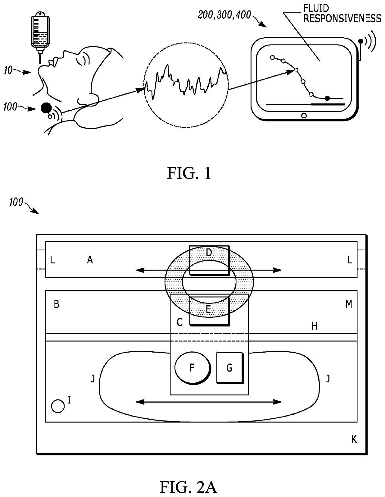
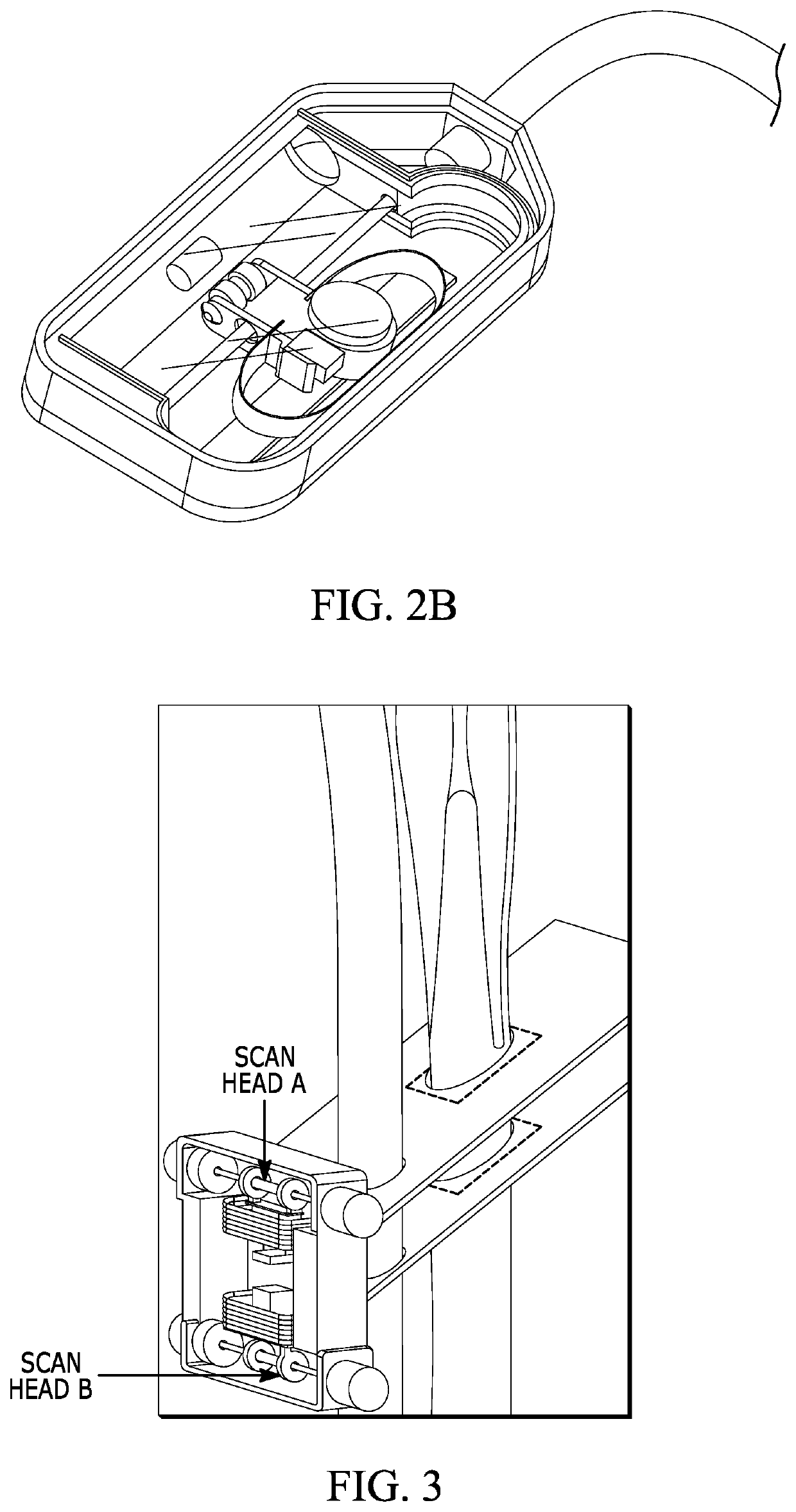
[0037]Provided are techniques for non-invasively, autonomously, and repeatedly measuring and recording changes of vessels over time using ultrasound technology. The ultrasound smart sensor system described herein performs fully automated or semi-automated (e.g., nurse-assisted) fluid status measurements similar to electrocardiograms, capnography, or pulse oximetry. An example sensor is a volume responsiveness (VR) sensor that can provide an automated, disposable, low-cost device that professionals can place on the side of a patient's neck, over the internal jugular vein (IA to obtain continuous and real-time assessments of a patient's fluid status. Generally speaking, the VR sensor is first applied to the patient's neck by a clinician, and subsequently, algorithms autonomously extract measurements obtained from the sensor at pre-programmed discrete intervals. A monitor automates this measurement process and records the resulting trends for use by medical professionals. In some examp...


PUM


Abstract

A fully automated ultrasound apparatus includes a sensor or probe which can be initially manually attached to a side of the neck of a patient, an ultrasound interface to control the sensor and periodically acquire raw ultrasound data, a signal and image processing system to autonomously convert the raw ultrasound data into a measurement that is useful to physicians, and a display to relay the current measurements and measurement history to provide data trends. The sensor can include one or more ultrasound transducers built into a housing. A disposable component can serve to secure the sensor to the neck of the patient and to provide a coupling medium between the sensor and the skin of the patient.

Description

CROSS-REFERENCE TO RELATED APPLICATION[0001]This application claims the priority benefit under 35 U.S.C. § 119(e) of U.S. Provisional Application No. 62 / 504,199, filed May 10, 2017, entitled “Automated Ultrasound Apparatus and Methods to Non-Invasively Monitor Fluid Responsiveness,” the disclosure of which is incorporated herein by reference in its entirety.STATEMENT REGARDING FEDERALLY SPONSORED RESEARCH OR DEVELOPMENT[0002]This invention was made with government support under 1600236 awarded by the National Science Foundation Innovation Corps. The government has certain rights in the invention.FIELD OF THE DISCLOSURE[0003]The present disclosure relates generally to medical devices and, more particularly, to techniques for utilizing ultrasound imaging to autonomously monitor changes in a vessel, such as geometric changes.BACKGROUND[0004]The background description provided herein is for the purpose of generally presenting the context of the disclosure. Work of the presently named in...

Claims


Application Information

Patent Timeline
IPC IPC(8): A61B8/08A61B8/00A61B8/06A61B8/02
CPCA61B8/02A61B8/5223A61B8/4236A61B8/5276A61B8/5207A61B8/4254A61B8/06A61B8/085A61B8/4461A61B8/4477A61B8/461A61B8/488A61B8/5246A61B8/5253A61B5/0095A61B5/02A61B5/6832A61B5/01A61B5/02416A61B5/026A61B5/0533A61B5/11A61B5/14542A61B5/6822A61B2562/04A61B2562/06A61B5/7257G16H50/30A61B5/327A61B5/318
Inventor KRUGER, GRANTBECKER, TORBEN K.THEYYUNNI, NIKHILKESSLER, ROSSPLUMMER, BRADLEYTAFOYA, MATTHEWTAFOYA, CHELSEAHARTE, STEVEN E.
Owner RGT UNIV OF MICHIGAN
Why Eureka
Try Eureka