Use of dendritic cells expressing foxp3 for diagnosis or treatment of cancer

Inactive Publication Date: 2020-06-11
RES & BUSINESS FOUNDATION SUNGKYUNKWAN UNIV
View PDF0 Cites 0 Cited by
  • Summary
  • Abstract
  • Description
  • Claims
  • Application Information

AI Technical Summary

Benefits of technology

This patent describes the use of dendritic cells that express a protein called Foxp3 and CD8+ Treg cells derived from these cells as markers for diagnosing and treating cancer. These cells can be found in tumors and the blood of cancer patients. This technology has applications in cancer research and therapy, and can help improve the monitoring of treatment outcomes.

Problems solved by technology

However, little is known about immune cells other than T cells, such as dendritic cells, in relation to Foxp3 expression and the medical utility thereof.

Method used


Image

  • Use of dendritic cells expressing foxp3 for diagnosis or treatment of cancer
  • Use of dendritic cells expressing foxp3 for diagnosis or treatment of cancer
  • Use of dendritic cells expressing foxp3 for diagnosis or treatment of cancer

Examples


example 1

Measurement of Blood fxDC in Tumor Patients

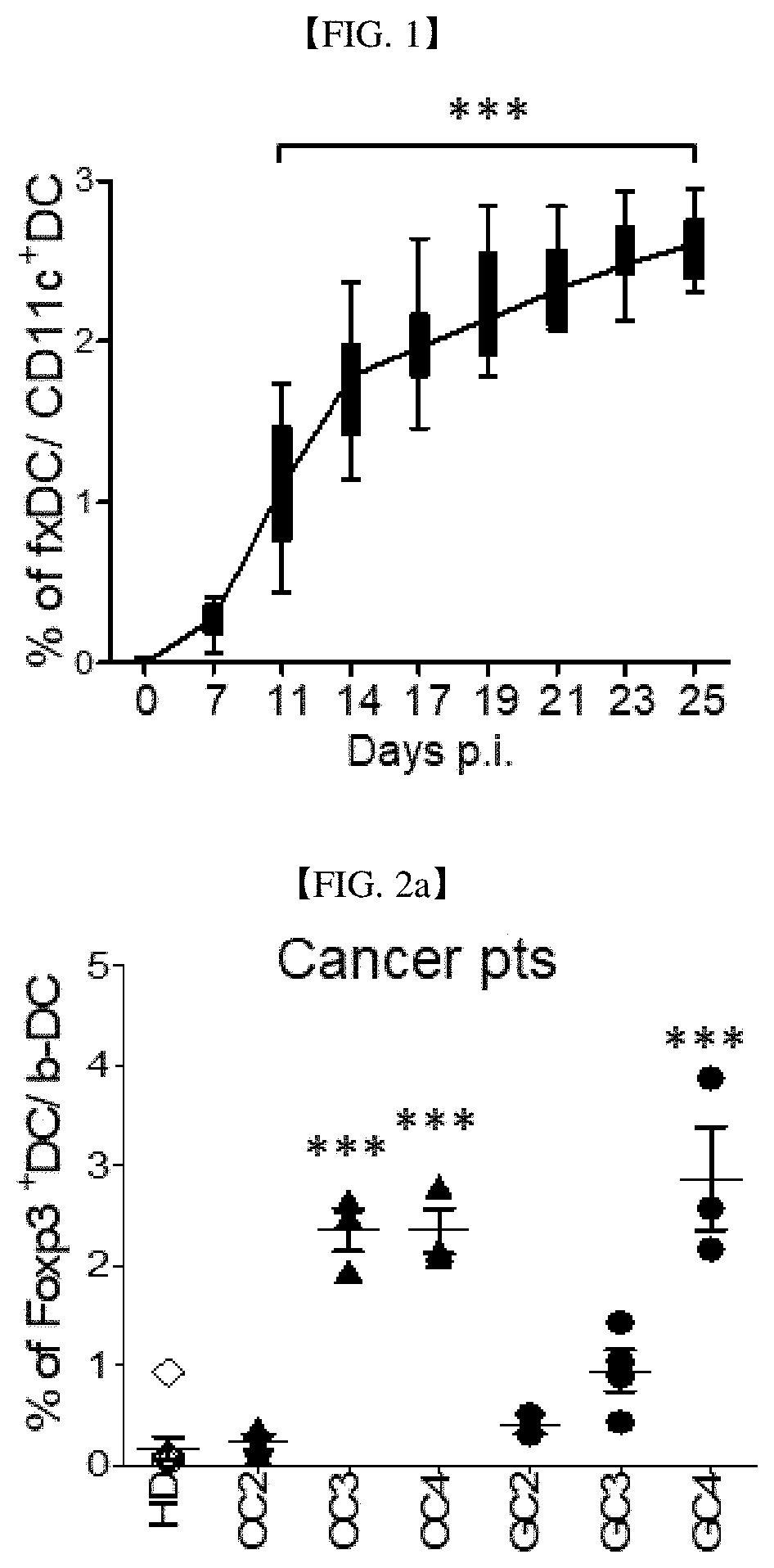
[0093]In mouse tumor models constructed by injecting EL4 lymphoma into mice (see Reference Example 2), orbital blood (ocular blood) collection was performed every three days from day 7 after tumor cell transplantation, followed by measuring Foxp3-expressing dendritic cells (expressed as fxDC or Foxp3+ DC) in the blood (see Reference Examples 4 and 5). The results are depicted in FIG. 1. FIG. 1 shows results of monitoring fxDC populations in the blood of Foxp3GFP mice during tumor growth, wherein fxDC was estimated at each time point for blood collected from the ocular veins of the mice in each group (n=30 for three groups, 10 mice per group). As shown in FIG. 1, the percentage of fxDC in the blood of the tumor mouse models appears to increase with tumor growth.

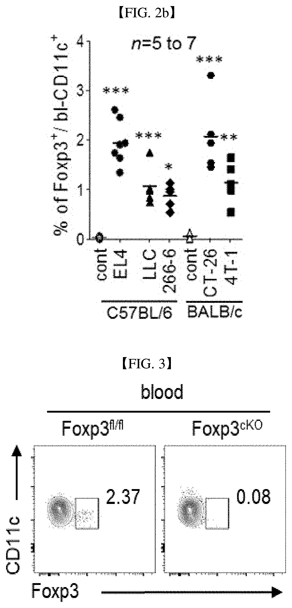
[0094]In addition, measurement was made of fxDC in blood DC (b-DC) (see Reference Examples 4 and 5) from healthy donors (HD) and cancer patients (glioblastoma (GBM, stages 3 and 4), c...

example 2

Assay for Tumor Growth Inhibition by fxDC Inhibition

[0096]To investigate the effect of fxDC on tumor growth, first, DC-specific Foxp3-knockout mice (CD11c-Cre×Foxp3fl / fl: hereinafter referred to Foxp3cKO) were constructed (see Reference Example 1), followed by injecting tumor cells thereto to prepare tumor mice before measurement of blood fxDC (see Reference Examples 4 and 5). The results are depicted in FIG. 3. As can be seen in FIG. 3, fxDC was depleted from the blood of Foxp3cKO mice.

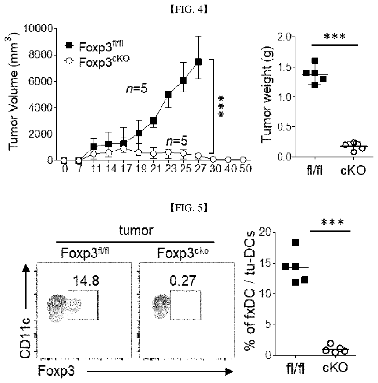
[0097]From seven days after injection of EL4 lymphoma tumor cells (5×10⁵ cells) thereto, wild-type mice (Foxp3fl / fl; TB mouse in which Foxp3 had not been knocked out) and Foxp3cKO mice were monitored every three days for tumor growth. The results are depicted in FIG. 4. FIG. 4 shows plots of tumor volumes vs. times (left) and tumor weights on day 23 after tumor transplantation (right). As shown in FIG. 4, wild-type mice (Foxp3fl / fl) gradually increased in tumor size whereas tumors in the fxDC-deplete...

example 3

Assay for Increase of CD8+ T Cells and Cytotoxicity Against Tumor Cells by fxDC Inhibition

[0100]CD8+ T (Tc1) cells play a crucial role in anti-cancer immunity and directly induce the apoptosis of tumor cells (cytotoxic CD8+T-cell). CD8+ T cells in the tumor of fxDC-depleted Foxp3cKO mice amounted to about 35.6%, which was observed to be a great increase over the proportion (about 16.3%) of CD8+ T cells in wild-type mice (Foxp3fl / fl). Among CD8+ T cells in tumor tissues of fxDC-depleted Foxp3cKO mice, proportions of IFN-gamma-expressing CD8+ T cells (IFN-gamma+ CD8+ T cells; cytotoxic CD8+ T-cells) were measured, and the results are depicted in FIG. 7. As shown in FIG. 7, the proportion of the cytotoxic CD8+ T-cells in fxDC-depleted Foxp3cKO mice was 2.5 times as large as that in wild-type mice (Foxp3fl / fl). The results suggest the regulatory effect of fxDC on cytotoxic CD8+ T-cells (upregulation of cytotoxic CD8+ T-cell by fxDC depletion).

[0101]Investigation was made to see whether ...


PUM


Abstract

Provided is a use of at least one selected from the group consisting of Forkhead box P3 (Foxp3)-expressing dendritic cells and cluster of differentiation 8 (CD8)-positive regulatory T cells as a target for cancer therapy and / or as a marker for cancer diagnosis.

Description

TECHNICAL FIELD[0001]Provided is a use of at least one selected from the group consisting of Forkhead box P3 (Foxp3)-expressing dendritic cells and cluster of differentiation 8 (CD8)-positive regulatory T cells as a target for cancer therapy and / or as a marker for cancer diagnosis.BACKGROUND ART[0002]Dendritic cells (DCs) are antigen-presenting cells (APCs) of the mammalian immune system, which act as important messengers between the innate and the adaptive immune systems.[0003]Forkhead box P3 (Foxp3) is a transcriptional regulatory factor known to be involved in the development and function of regulatory T cells (Treg) (Hori, S., Nomura, T. & Sakaguchi, S. Control of regulatory T cell development by the transcription factor Foxp3. Science 299, 1057-1061, doi:10.1126 / science.1079490 (2003)).[0004]However, little is known about immune cells other than T cells, such as dendritic cells, in relation to Foxp3 expression and the medical utility thereof.DETAILED DESCRIPTION OF THE INVENTIO...

Claims


Application Information

Patent Timeline
IPC IPC(8): C07K16/28A61K47/64A61P35/00
CPCC07K16/2866A61K47/642A61K38/00A61P35/00G01N33/50G01N33/68C07K16/18A61K39/4615A61K39/464499C12N5/0637A61K39/4621A61K2239/48A61K39/4622A61K39/4611A61K39/395G01N33/5011G01N33/5047G01N33/6872A61K2039/505G01N2333/70517C12N2502/1114C12N2502/1121
Inventor BAE, YONG SOOJEONG, YI DEULKANG, MYONG HO
Owner RES & BUSINESS FOUNDATION SUNGKYUNKWAN UNIV
Why Eureka
Try Eureka