Determining biomarkers from histopathology slide images

a biomarker and histopathology technology, applied in image enhancement, medical/anatomical pattern recognition, instruments, etc., can solve the problems of low 5-year survival rate, lack of resources, and limited ihc staining

Active Publication Date: 2020-08-13
TEMPUS LABS INC
View PDF9 Cites 76 Cited by
  • Summary
  • Abstract
  • Description
  • Claims
  • Application Information

AI Technical Summary

Benefits of technology

[0049]In accordance with another example, a computing device configured to identifying biomarkers in digital image of a Hematoxylin and eosin (H&E) stained slide of target tissue, the computing device comprises: one or more memories; and one or more processors configured to, receive the digital image; perform an image tiling process on the digital image by separating the digital image into a plurality of tile images, where each of the plurality of tile images contains a different portion of the digital image; apply the plurality of tile images to a multiscale deep learning framework comprising one or more trained deep learning multiscale classifier models, each trained deep learning multiscale classifier models being trained to classify a different tissue classification for each tile image and determine a tissue classification for each of the plurality of tile images, using the multiscale deep learning framework; identify cells within the digital image using a trained cell segmentation model; and from the tissue classification determined for each tile image and from the identified cells within the digital image, identify a predicted presence of one or more biomarkers associated with the digital image.
[0050]In accordance with another example, a computing device configured to identifying biomarkers in digital image of a Hematoxylin and eosin (H&E) stained slide of target tissue, the computing device comprises: one or more memories; and one or more processors configured to, receive a molecular training dataset for a plurality of training tissue samples, the molecular training dataset comprising RNA transcriptome counts from sequencing of a substantially similar sample associated with each training tissue sample; perform a clustering process on the molecular training dataset to identify one or more molecular data subsets each corresponding to a different biomarker; for each o

Problems solved by technology

NSCLC often responds poorly to standard of care chemoradiotherapy and has a high incidence of recurrence, resulting in low 5-year survival rates.
However, such IHC staining is often limited by insufficient tissue samples and, in some settings, a lack of resources.
However, neighboring sections of the image overlap, such that processing each section separately would create a high number of

Method used


Image

  • Determining biomarkers from histopathology slide images
  • Determining biomarkers from histopathology slide images
  • Determining biomarkers from histopathology slide images

Examples


Embodiment Construction

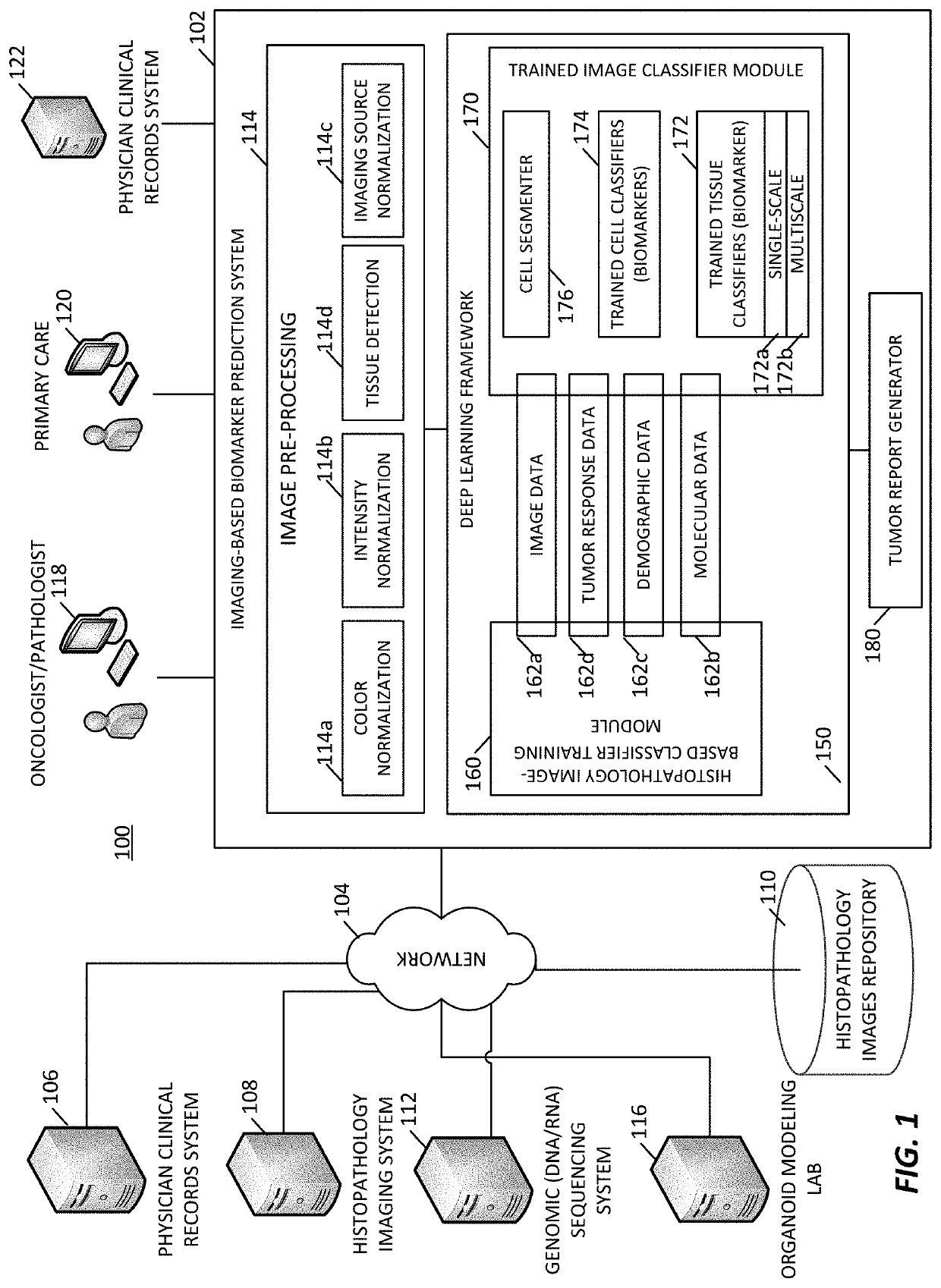
[0084]An imaging-based biomarker prediction system is formed of a deep learning framework configured and trained to directly learn from histopathology slides and predict the presence of biomarkers in medical images. The deep learning frameworks may be configured and trained to analyze medical images and identify biomarkers that indicate the presence of a tumor, a tumor state / condition, or information about a tumor of the tissue sample.

[0085]In an implementation, a cloud-based deep learning framework is used for medical image analysis. Deep learning algorithms automatically learn sophisticated imaging features for enhanced diagnosis, prognosis, treatment indication, and treatment response prediction. In examples, the deep learning frameworks are able to directly connect to cloud storage and leverage resources on cloud platforms for efficient deep learning algorithm training, comparison, and deployment.

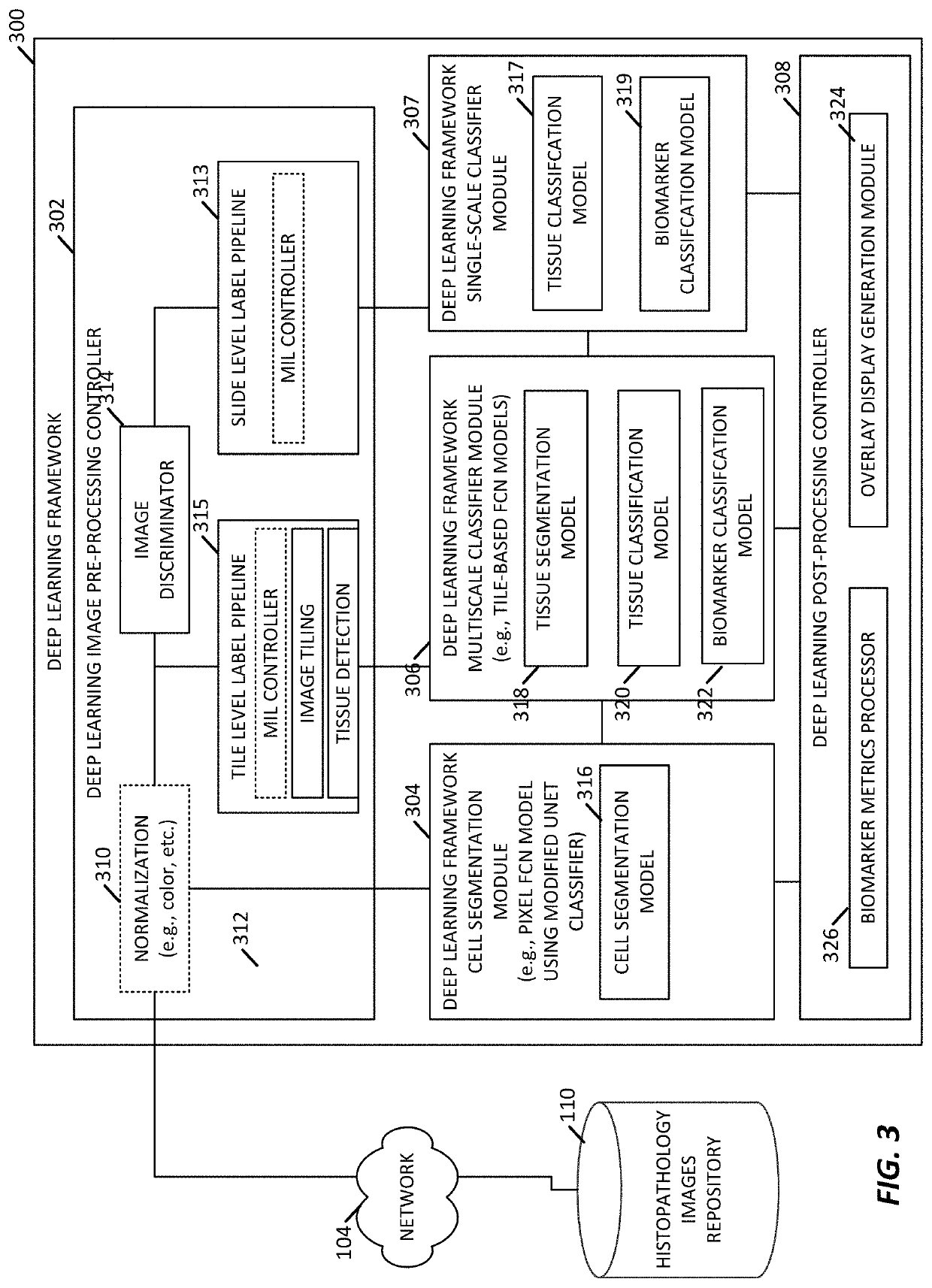
[0086]In some examples, the deep learning frameworks include a multiscale configura...


PUM


Abstract

A generalizable and interpretable deep learning model for predicting biomarker status and biomarker metrics from histopathology slide images is provided.

Description

CROSS-REFERENCE TO RELATED APPLICATIONS[0001]This application is a Continuation-in-Part of U.S. application Ser. No. 16 / 732,242, filed on Dec. 31, 2019, which claims priority to U.S. Provisional Application Ser. No. 62 / 787,047, filed Dec. 31, 2018, and is a Continuation-in-Part of U.S. application Ser. No. 16 / 412,362, filed on May 14, 2019, which claims priority to U.S. Provisional Application No. 62 / 671,300 filed May 14, 2018, and claims priority to U.S. Provisional Application Ser. No. 62 / 824,039, filed on Mar. 26, 2019, U.S. Provisional Application Ser. No. 62 / 889,521, filed Aug. 20, 2019 and to U.S. Provisional Application Ser. No. 62 / 983,524, filed on Feb. 28, 2020, the entire disclosures of each of which are hereby expressly incorporated by reference herein.FIELD OF THE INVENTION[0002]The present disclosure relates to examining digital images to detect, quantify, and / or characterize cancer-related biomarker(s) and, more particularly, to detect, quantify, and / or characterize su...

Claims


Application Information

Patent Timeline
IPC IPC(8): G06T7/00G06K9/62G06T7/11G06T11/00G06T1/20G06V10/44G06V10/764
CPCG06T7/11G06T1/20G06T11/00G06T7/0012G06T2207/30024G06K9/628G06K9/6217G06T2207/30096G06T2207/20081G06T2207/10056G06T2207/10024G06T2207/20084G06N3/084G06V10/44G06V2201/03G06V10/82G06V10/764G06N3/045G06F18/21G06F18/2431
Inventor YIP, STEPHENHO, IRVINSHA, LINGDAOOSINSKI, BOLESLAWKHAN, ALY AZEEMKRUGER, ANDREW J.CARLSON, MICHAELGREENWALD, ABELWILLIS, CALEB
Owner TEMPUS LABS INC
Why Eureka
Try Eureka