Semi-automated imaging reconstruction for orbital fracture repair

a technology of orbital fracture and imaging reconstruction, which is applied in the field of semi-automated imaging reconstruction for orbital fracture repair, can solve the problems of poor signal contrast of orbital bones from adjacent tissues (e.g. paranasal sinuses) in medical images, and the difficulty in identifying and delineating the complete extent of orbital fracture in these images, so as to achieve the effect of enhancing contras

Active Publication Date: 2020-08-20
THE CHINESE UNIVERSITY OF HONG KONG
View PDF0 Cites 6 Cited by
  • Summary
  • Abstract
  • Description
  • Claims
  • Application Information

AI Technical Summary

Benefits of technology

[0004]The image processing system can be trained to detect the orbital fracture using multiple orbital images with and without fractures. For example, the image processing system can be trained to detect any discontinuity of orbital bone structures and / or prolapse of orbital tissue into surrounding sinuses as indications of the orbital fracture. Generating the three-dimensional reconstruction model may include, for each of the two-dimensional images in which the orbital fracture is detected, adaptively enhancing contrast along the extracted region of interest (fractured areas), converting greyscale color into a binary black and white, and / or segmenting irregular boundaries using an active contour algorithm. Generating the three-dimensional reconstruction model may also include applying a marching cubes algorithm to generate an isosurface for the three-dimensional model.

Problems solved by technology

Existing techniques for surgical reconstruction of the fractured orbit poses challenges that are different from the rest of the body.
As orbital bones are among the thinnest in the human body, signal contrast of the orbital bones from the adjacent tissues (e.g. paranasal sinuses) can be poor in medical images.
Thus, there is an inherent difficulty in identifying and delineating the complete extent of orbital fracture in these images.
This process of segmentation is time-consuming and requires intensive input from both clinicians and engineers.
Detection of bone fractures over these commonly fractured regions is particularly difficult.
On the other hand, current process of manual segmentation and reconstruction of any 3D model from multiple two-dimensional (2D) scan images is also extremely labor-intensive.
This remains the rate-limiting step in generating a 3D model for operative planning in orbital fracture repair.

Method used


Image

  • Semi-automated imaging reconstruction for orbital fracture repair
  • Semi-automated imaging reconstruction for orbital fracture repair
  • Semi-automated imaging reconstruction for orbital fracture repair

Examples


Embodiment Construction

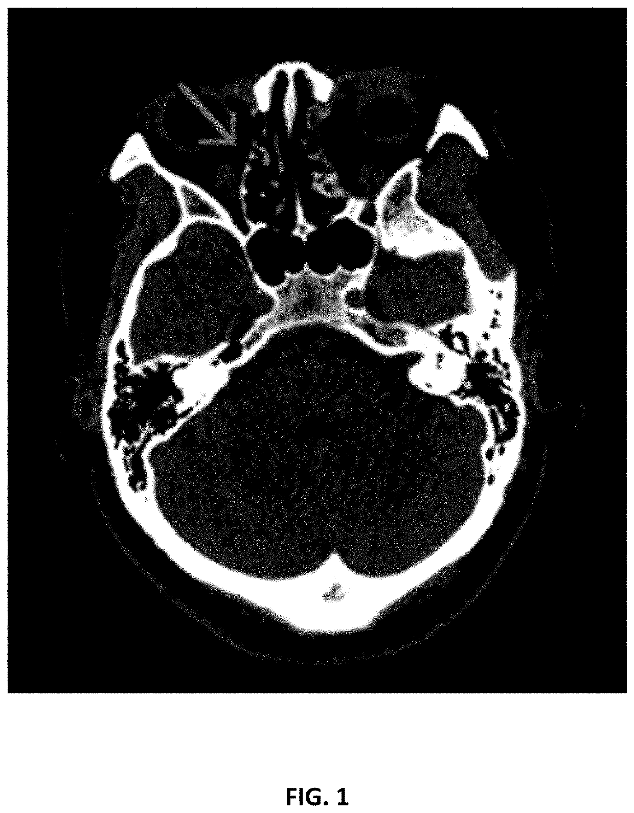
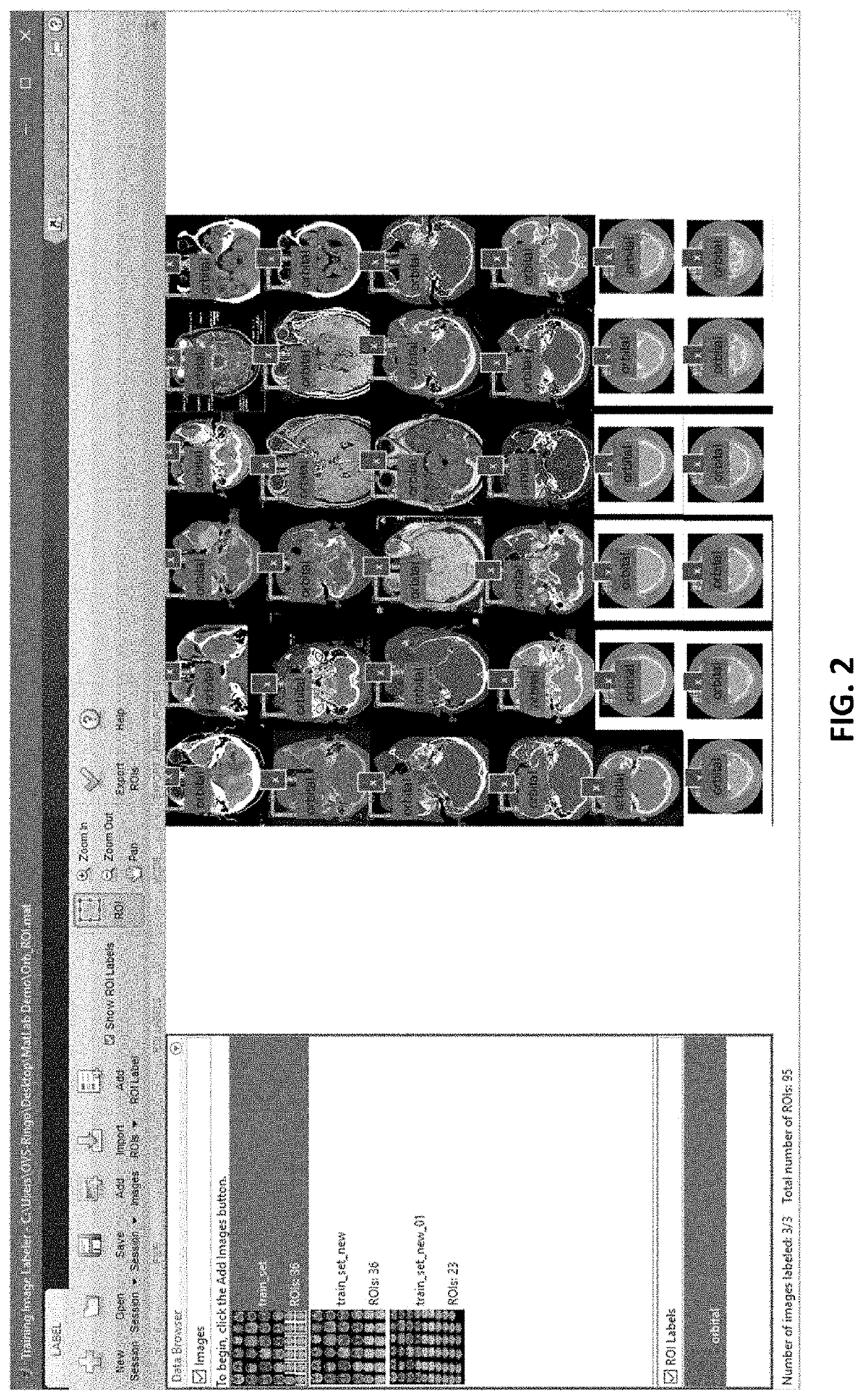
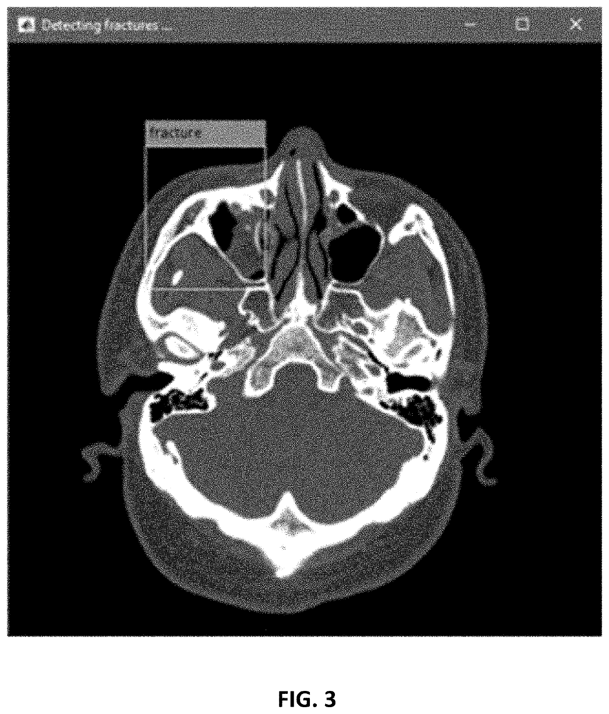
[0019]Various aspects of the present invention relate to the design of patient-specific orbital implants and instruments, and the process involved in production of such implants. In particular, techniques for 3D modelling anatomical models for reconstruction of orbital fracture using image-processing are described herein. The image-processing algorithms are automated as much as possible to shorten the time from clinical imaging to surgical implantation. These processes include automated detection of orbital fracture from medical images (e.g., CT images), identifying and cropping the region of interest (ROI) for further processing, segmentation of the orbital bony ROI using image processing techniques, reconstruction of the 3D model of the pre-fractured orbit from these segmented images, and output of the 3D model data for fabrication of the orbital models and instrument. The 3D model is generated in stereolithographical file for 3D printing of the orbital molds and instruments.

[0020...


PUM

PropertyMeasurementUnit
thickaaaaaaaaaa
thickaaaaaaaaaa
image processingaaaaaaaaaa

Abstract

Techniques for fabrication of implant material for the reconstruction of fractured eye orbit may include using an image processing system to analyze a set of two-dimensional images representing a three-dimensional scan of a skull of a patient, automatically detect an orbital fracture in the skull based on the set of two-dimensional images, and identify which / both of the two eye orbits containing any orbital fracture. The techniques may further include, for each of the two-dimensional images in which the orbital fracture is detected, determining a region of interest, and extracting the region of interest. The techniques may further include generating a three-dimensional reconstruction model for the fractured eye orbit, and outputting model data for generating an implant mold for the fractured eye orbit.

Description

CROSS-REFERENCES TO RELATED APPLICATIONS[0001]This application claims the benefit of priority to U.S. Provisional Application No. 62 / 807,480, filed Feb. 19, 2019, the entire contents of which is incorporated herein by reference.BACKGROUND[0002]Existing techniques for surgical reconstruction of the fractured orbit poses challenges that are different from the rest of the body. As orbital bones are among the thinnest in the human body, signal contrast of the orbital bones from the adjacent tissues (e.g. paranasal sinuses) can be poor in medical images. Thus, there is an inherent difficulty in identifying and delineating the complete extent of orbital fracture in these images. Therefore, reconstruction of three-dimensional (3D) model of the orbital region for reconstructive purposes has exclusively been done manually. This process of segmentation is time-consuming and requires intensive input from both clinicians and engineers. The most widely used medical imaging modality in orbital fr...

Claims


Application Information

Patent Timeline
Patent Type & Authority Applications(United States)
IPC IPC(8): A61F2/30G06T7/00G06T17/00G06K9/62G06T5/00G06T7/149A61B17/02A61F2/28
CPCG06T2207/30008A61F2/2875A61F2002/30985A61F2/30942G06K9/6228G06T2210/41A61B2017/0042G06T7/0012A61F2002/30957G06T2207/30041G06T17/00G06T2207/20116G06T7/149A61F2002/30948G06T5/008A61F2002/2878G06K9/6256A61B17/0231A61B2017/00526A61F2002/30952G06T2207/20081G06K2209/055A61B17/02A61B2034/108A61B34/10G06T7/11G06T2207/10081A61F2002/30943A61F2002/30962G06V2201/033G06F18/211G06F18/214
Inventor CHONG, KELVIN KAM-LUNGCHUI, CHUN SINGCHEUNG, WING HING RINGO
Owner THE CHINESE UNIVERSITY OF HONG KONG
Why Eureka
Try Eureka