Methods of isolating t cells having antigenic specificity for a p53 cancer-specific mutation

Pending Publication Date: 2020-10-08
US DEPT OF HEALTH & HUMAN SERVICES
View PDF2 Cites 3 Cited by
  • Summary
  • Abstract
  • Description
  • Claims
  • Application Information

AI Technical Summary

Benefits of technology

[0008]Another embodiment of the invention provides method of preparing a population of T cells that have antigenic specificity for a mutated p53 amino acid sequence encoded by a cancer-specific p53 mutation, the method comprising: isolating T cells acco

Problems solved by technology

Nevertheless, obstacles to the successful use of ACT for the widespread treatment of cancer and other diseases remain.
For

Method used


Image

  • Methods of isolating t cells having antigenic specificity for a p53 cancer-specific mutation
  • Methods of isolating t cells having antigenic specificity for a p53 cancer-specific mutation
  • Methods of isolating t cells having antigenic specificity for a p53 cancer-specific mutation

Examples


example 1

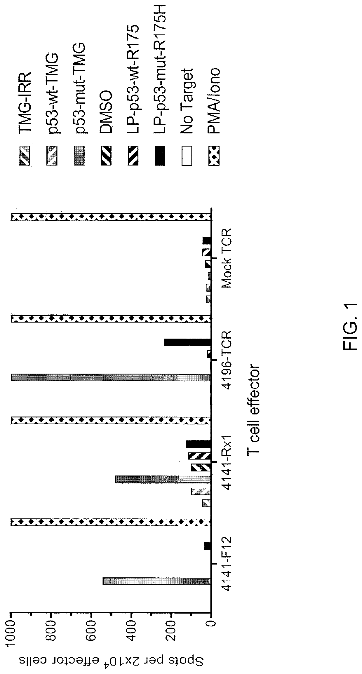
[0123]This example demonstrates the identification of anti-mutated p53 T cells in Patient 4141 by co-culturing autologous APCs induced to express mutated p53 within autologous T cells (“p53 hotspot mutation universal screening”). This example also demonstrates the isolation and specific reactivity of a TCR from patient 4141.

[0124]Experiments were carried out as described for FIGS. 1 and 45-48 for Patient 4141. TIL fragment F12 and infusion bag TIL (Rx1) from patient 4141 and p53-R175H-specific TCR or mock transduced T cells from patient 4196 were used as effectors. Co-cultures with T cell effectors and HLA-A*02:01 APCs (autologous to patient 4141) were either (1) electroporated with TMGs composed of irrelevant, WT p53, or mutated p53 sequences or (2) pulsed with peptide vehicle (DMSO) or purified (>95% by HPLC) 25 amino acid peptides composed of WT p53-R175 sequence or mutated p53-R175H sequence. T cells only (no target) was negative control and PMA and Iono was positive control (la...

example 2

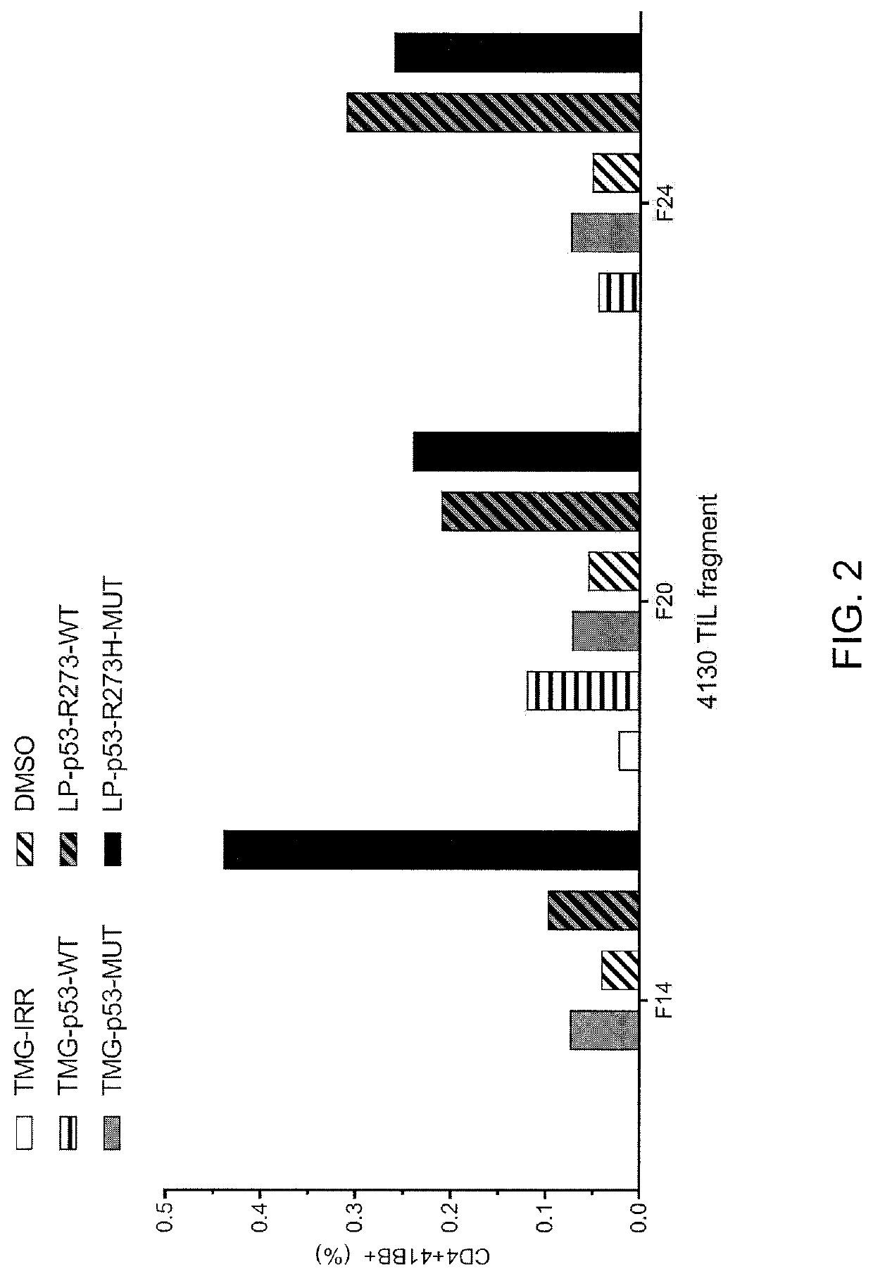
[0132]This example demonstrates the identification of anti-mutated p53 T cells in Patient 4130 by co-culturing autologous APCs induced to express mutated p53 within autologous T cells (“p53 hotspot mutation universal screening”).

[0133]Experiments were carried out as described for FIG. 2 for Patient 4130. TIL fragments (F14, F20 and F24) from patient 4130 were co-cultured with autologous APCs (1) electroporated with TMGs composed of irrelevant, WT p53 or mutated p53 sequences or (2) pulsed with peptide vehicle (DMSO) or purified (>95% by HPLC) 25 amino acid peptides composed of WT p53-R273 sequence or mutated p53-R273H sequence. Co-cultures were performed overnight at 37° C. Expression of 4-1BB was evaluated by flow cytometry after gating for lymphocytes→living cells (PI negative)→CD3+ (T cells). The results are shown in FIG. 2.

example 3

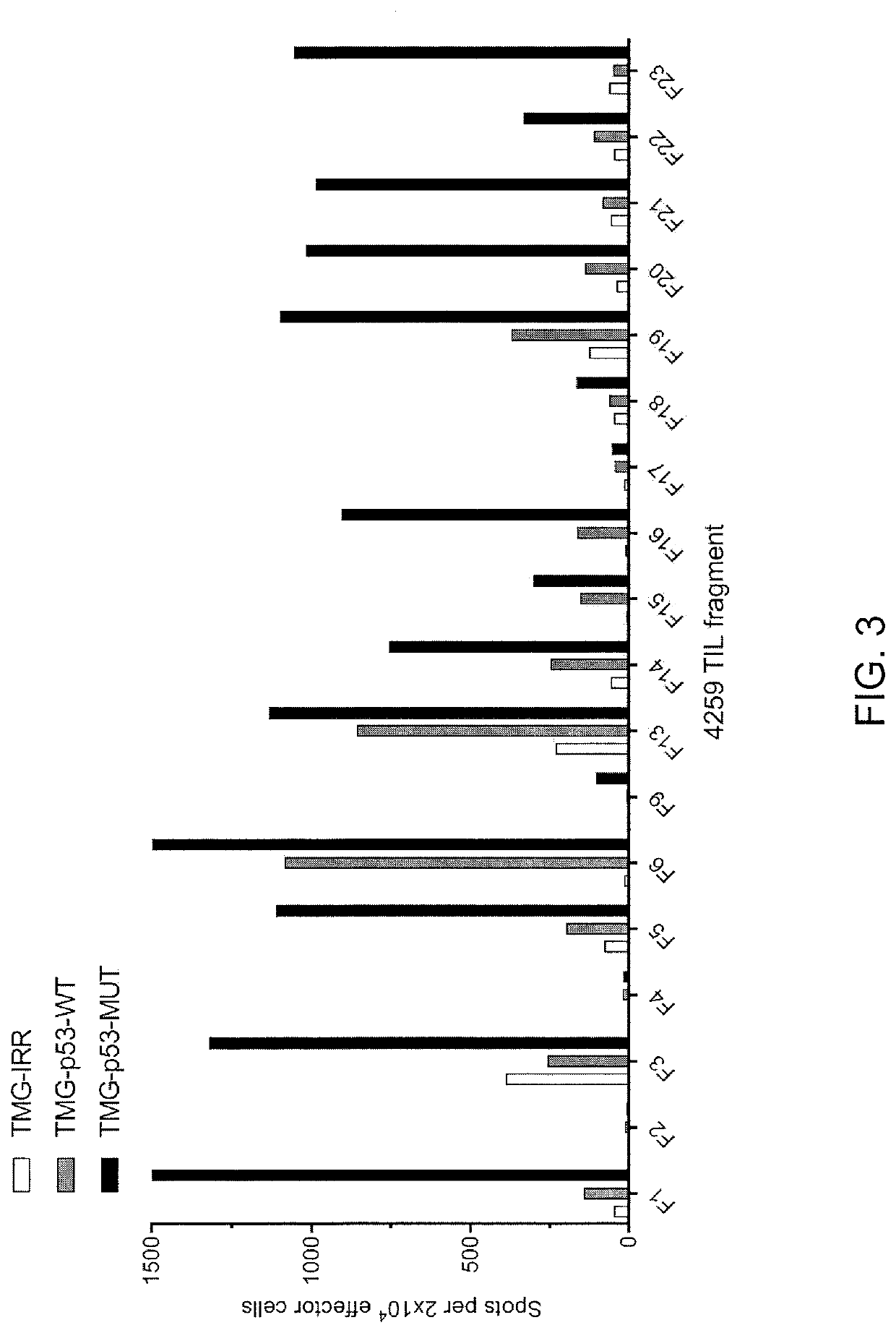
[0134]This example demonstrates the identification of anti-mutated p53 T cells in Patient 4259 by co-culturing autologous APCs induced to express mutated p53 within autologous T cells (“p53 hotspot mutation universal screening”). This example also demonstrates the isolation and specific reactivity of a TCR isolated from patient 4259.

[0135]Experiments were carried out as described for FIGS. 3-8 and 49-53 for Patient 4259. TIL fragments (n=18) from patient 4259 were co-cultured with autologous APCs electroporated with TMG composed of irrelevant, WT p53 or mutated p53 sequences. Co-cultures were performed overnight at 37° C. Secretion of IFN-γ was evaluated using ELISPOT assay. The results are shown in FIG. 3. Expression of 4-1BB was evaluated by flow cytometry after gating for lymphocytes→living cells (PI negative)→CD3+ (T cells). The results are shown in FIGS. 4-5.

[0136]TIL fragments (n=18) from patient 4259 were co-cultured with autologous APCs pulsed with peptide vehicle (DMSO) or ...


PUM

PropertyMeasurementUnit
Volumeaaaaaaaaaa
Molar densityaaaaaaaaaa
Fractionaaaaaaaaaa

Abstract

Disclosed are methods of isolating T cells having antigenic specificity for a mutated p53 amino acid sequence encoded by a cancer-specific p53 mutation, the method comprising: inducing autologous APCs of the patient to present the mutated p53 amino acid sequence; co-culturing autologous T cells of the patient with the autologous APCs that present the mutated p53 amino acid sequence; and selecting the autologous T cells. Also disclosed are related methods of preparing a population of cells, populations of cells, pharmaceutical compositions, and methods of treating or preventing cancer.

Description

CROSS-REFERENCE TO RELATED APPLICATION[0001]This patent application claims the benefit of U.S. Provisional Patent Application No. 62 / 565,464, filed Sep. 29, 2017, which is incorporated by reference in its entirety herein.STATEMENT REGARDING FEDERALLY SPONSORED RESEARCH AND DEVELOPMENT[0002]This invention was made with Government support under project number ZIABC010984 by the National Institutes of Health, National Cancer Institute. The Government has certain rights in the invention.INCORPORATION-BY-REFERENCE OF MATERIAL SUBMITTED ELECTRONICALLY[0003]Incorporated by reference in its entirety herein is a computer-readable nucleotide / amino acid sequence listing submitted concurrently herewith and identified as follows: One 687,616 Byte ASCII (Text) file named “740175 ST25.txt,” dated Sep. 14, 2018.BACKGROUND OF THE INVENTION[0004]Adoptive cell therapy (ACT) can produce positive clinical responses in some cancer patients. Nevertheless, obstacles to the successful use of ACT for the wid...

Claims


Application Information

Patent Timeline
IPC IPC(8): A61K35/17C07K14/47C12N5/0783
CPCC07K14/4746C12N5/0636A61K35/17C12N2502/1121A61K39/001151A61P35/00C07K14/47
Inventor DENIGER, DREW C.ROSENBERG, STEVEN A.MALEKZADEH, PARISA
Owner US DEPT OF HEALTH & HUMAN SERVICES
Why Eureka
Try Eureka