Combined assessment of morphological and perivascular disease markers

a morphological and perivascular disease technology, applied in the field of combined assessment of morphological and perivascular disease markers, can solve the problems of complex structure of tissues both inside and outside the vessel wall, difficult to interpret raw pixel reconstructed intensity values using simplistic thresholding operators,

Active Publication Date: 2021-02-11
ELUCID BIOIMAGING INC
View PDF0 Cites 33 Cited by
  • Summary
  • Abstract
  • Description
  • Claims
  • Application Information

AI Technical Summary

Benefits of technology

[0019]In some embodiments, segmenting the medical imaging data further comprises segmenting the medical imaging data into an outer wall boundary. In some embodiments, the analyzer module is configured to partition a lumen and an outer wall based on the segmented lumen boundary and outer wall boundary into one or more vessel boundaries. In some embodiments, the biological properties include calcified regions, LRNC regions, intra-plaque regions, matrix regions, or any combination thereof.

Problems solved by technology

Currently difficulties with the recent progress can include difficulty with interpreting raw pixel reconstructed intensity values using simplistic thresholding operators.
Additionally, heterogeneity of tissues both within and outside the vessel wall present classification and measurement challenges unless processed effectively.

Method used


Image

  • Combined assessment of morphological and perivascular disease markers
  • Combined assessment of morphological and perivascular disease markers
  • Combined assessment of morphological and perivascular disease markers

Examples


Embodiment Construction

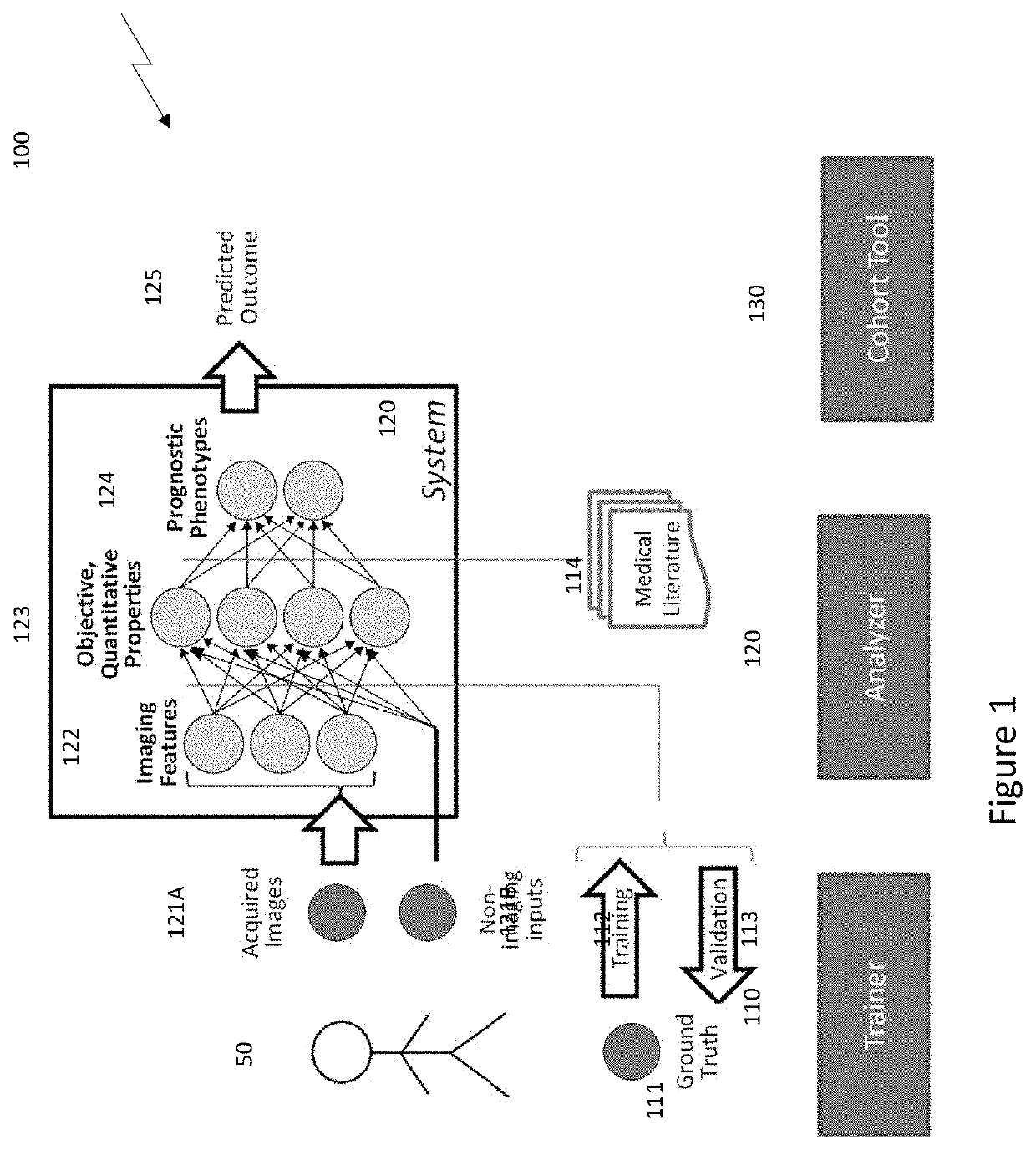
[0033]FIG. 1 is a diagram of a system for determining and characterizing a medical condition by implementing a hierarchical analytics framework, according to some embodiments of the invention. The system 100 can include a trainer module 110, an analyzer module 120 and a cohort tool module 120.

[0034]The analyzer module 120 can include a hierarchical analytics framework which can identify and / or quantify biological properties / analytes 130 based on medical imaging data (e.g., medical imaging data of patient). The medical imaging data can include (i) imaging features 122 from one or more acquired images 121A of a patient 50 and / or (ii) non-imaging input data 121B for a patient 50. The analyzer module 120 can identify and / or characterize one or more pathologies (e.g., prognostic phenotypes) 124 based on the quantified biological properties / analytes 123. The analyzer module 120 can operate independent of ground truth and / or validation references by implementing one or more pre-trained alg...


PUM


Abstract

A system including a hierarchical analytics framework that can utilize a first set of machine learned algorithms to identify and quantify a set of biological properties utilizing medical imaging data is provided. System can segment the medical imaging data based on the quantified biological properties to delineate existence of perivascular adipose tissue. The system can also segment the medical imaging data based on the quantified biological properties to determine a lumen boundary and/or determine a cap thickness based on a minimum distance between the lumen boundary and LRNC regions.

Description

CROSS REFERENCE TO RELATED APPLICATIONS[0001]This application claims the benefit of and priority to U.S. provisional patent application No. 62 / 882,881, filed on Aug. 5, 2019, the entire contents of which are incorporated herein by reference in its entirety and owned by the assignee of the instant application.GOVERNMENT RIGHTS[0002]This work was supported in part by NIH award ID HL 126224. The government may have certain rights in the invention.FIELD OF THE INVENTION[0003]The invention relates to computer-aided phenotyping (CAP) of disease which can include applying computerized image analysis and / or data fusion algorithms to patient data. In particular, the invention relates to quantitative imaging and analytics for elucidating the disease process of atherosclerosis, including delineating perivascular adipose tissue and / or determining the thickness of tissues between the lipid-rich necrotic core (LRNC) and the lumen (“cap thickness”) of a blood vessel.BACKGROUND OF THE INVENTION[000...

Claims


Application Information

Patent Timeline
Patent Type & Authority Applications(United States)
IPC IPC(8): G06T7/00G06T7/11A61B5/02A61B6/00A61B8/08
CPCG06T7/0012G06T7/11G06T2207/30101A61B6/504A61B8/0891A61B5/02007G06T2207/10081G06T2207/10088G06T2207/20081G06T2207/20084A61B6/032A61B6/5217A61B8/08G16H50/20A61B5/7264A61B5/4848G16H30/40G06T7/155
Inventor BUCKLER, ANDREW J.
Owner ELUCID BIOIMAGING INC
Why Eureka
Try Eureka