System and method for high-resolution, high-speed capsule endomicroscopy

a capsule endomicroscope and high-speed technology, applied in the field of high-resolution, high-speed capsule endomicroscope, can solve the problems of reducing the lifetime of water-filled secm capsules, affecting optimal performance, and difficult to achieve diffraction-limited optical resolution

Pending Publication Date: 2021-03-04
THE GENERAL HOSPITAL CORP
View PDF6 Cites 0 Cited by
  • Summary
  • Abstract
  • Description
  • Claims
  • Application Information

AI Technical Summary

Benefits of technology

[0005]Thus, a new SECM probe design was sought which does not require filling the probe w

Problems solved by technology

However, in prior designs asymmetric spherical aberrations caused at a probe surface (for example, arising from the cylindrical imaging window of an SECM capsule) have made it difficult to achieve diffraction-limited optical resolution.
However, filling a probe such as an SECM capsule with water poses a number of additional manufacturing requirements

Method used


Image

  • System and method for high-resolution, high-speed capsule endomicroscopy
  • System and method for high-resolution, high-speed capsule endomicroscopy
  • System and method for high-resolution, high-speed capsule endomicroscopy

Examples


Embodiment Construction

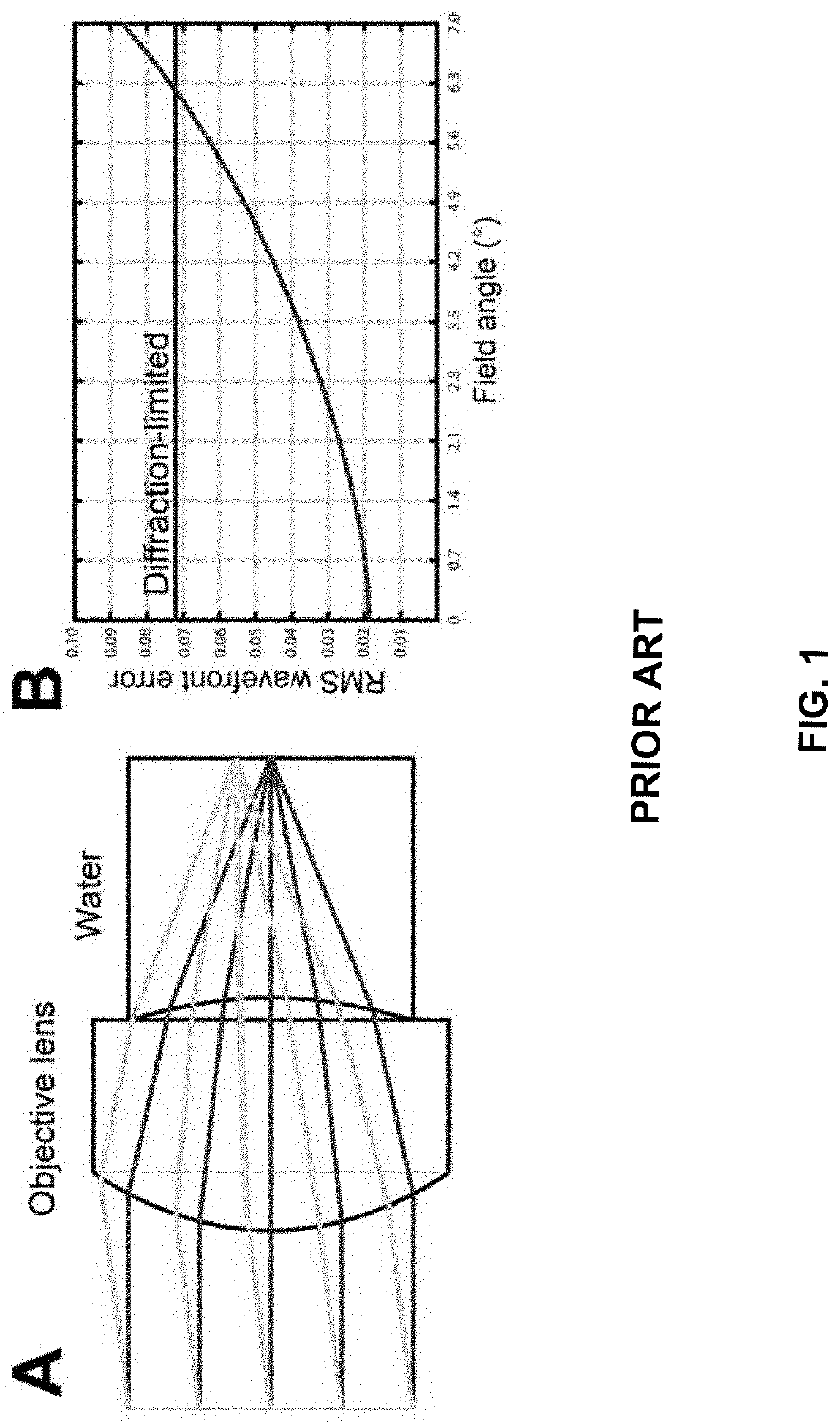
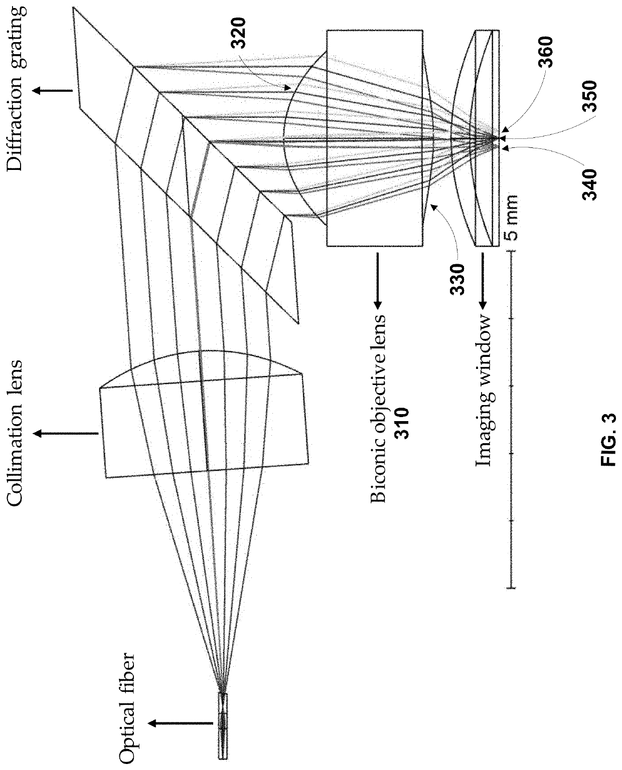
[0020]Disclosed herein are embodiments of an apparatus, method, and / or system for an SECM probe which includes an objective lens having a biconic surface and which as a consequence does not require water immersion or other corrective measures to be taken to achieve diffraction-limited or near diffraction-limited optical resolution.

[0021]Spectrally encoded confocal microscopy (SECM) is a miniature endomicroscopy technique that encodes physical locations on a specimen using wavelength to achieve fast imaging speeds. Briefly, a broadband or wavelength-swept light source is dispersed across a swath of a sample so that each wavelength or subgroups of wavelengths acts as a separate beam to illuminate a sample (FIG. 3), increasing the amount of data that can be collected from a sample in a single pass since this permits large amounts of data to be collected in parallel. While some pill-sized tethered SECM capsules have been developed to acquire cellular-level resolution images of the upper...


PUM


Abstract

A probe for performing endomicroscopy, including: a light source; a waveguide coupled to the light source; a diffraction grating, the waveguide directing light from the light source to the diffraction grating; and a lens having a first aspheric surface and a second biconic surface, diffracted light from the diffraction grating being directed into the aspheric surface of the lens and being emitted from the biconic surface of the lens towards a transparent cylindrical surface of the probe.

Description

CROSS-REFERENCE TO RELATED APPLICATIONS[0001]This application is based on, claims the benefit of, and claims priority to U.S. Provisional Application No. 62 / 892,073, filed Aug. 27, 2019, which is hereby incorporated herein by reference in its entirety for all purposes.STATEMENT REGARDING FEDERALLY SPONSORED RESEARCH[0002]N / ABACKGROUND[0003]Spectrally encoded confocal microscopy (SECM) is a high-speed imaging technique that can provide 1-2 orders of magnitude faster imaging speed compared to video rate reflectance confocal microscopy, while enabling a high degree of probe miniaturization by replacing a mechanical scanning device with a diffraction grating. However, in prior designs asymmetric spherical aberrations caused at a probe surface (for example, arising from the cylindrical imaging window of an SECM capsule) have made it difficult to achieve diffraction-limited optical resolution. One solution to this has been to fill the gap between the objective lens and the imaging window ...

Claims


Application Information

Patent Timeline
IPC IPC(8): G02B5/18A61B1/00A61B5/00
CPCG02B5/1814A61B5/0068A61B1/00165A61B1/00188G02B21/0028G02B23/243G02B23/2476G02B5/1842A61B1/00096A61B1/00177A61B1/00172A61B1/041A61B1/0638A61B1/07G02B21/0064G02B23/2469
Inventor TEARNEY, GUILLERMO J.RYU, JIHEUN
Owner THE GENERAL HOSPITAL CORP
Why Eureka
Try Eureka