Method and device for obtaining vascular pressure difference

Pending Publication Date: 2021-08-26
PULSE MEDICAL IMAGING TECH (SHANGHAI) CO LTD
View PDF0 Cites 2 Cited by
  • Summary
  • Abstract
  • Description
  • Claims
  • Application Information

AI Technical Summary

Benefits of technology

The present invention provides a method for accurately calculating the pressure difference in blood vessels by introducing the concept of cross-sectional shape and considering the influence of factors such as plaque information. By obtaining a plane geometric image at each cross-sectional position of the target blood vessel and establishing a shape difference function, the blood vessel pressure difference can be accurately calculated and reflect the pressure changes at both ends of the blood vessel. This method can be used for other blood flow characteristic values and ensures accurate and reliable results.

Problems solved by technology

The deposition of lipids and carbohydrates in human blood on the vascular wall will form plaques on the vascular wall, which will then cause vascular stenosis; especially the vascular stenosis that occurs near the coronary artery of the heart will cause insufficient blood supply to the myocardium and induce coronary heart disease Diseases such as angina and angina pectoris pose a serious threat to human health.
Although conventional medical testing methods such as coronary angiography and CT can show the severity of coronary artery stenosis, they cannot accurately evaluate coronary ischemia.
Some patients will feel uncomfortable due to the injection of the drug, which makes the method of measuring the FFR value based on the pressure guide wire have greater limitations.
In addition, although the measurement of FFR guided by the pressure guide wire is an important indicator of coronary stenosis hemodynamics, the high cost of the pressure guide wire and the difficulty in the operation of the interventional vascular process have severely restricted the measurement of FFR value based on the pressure guide wire.
The results of FFR evaluation of coronary stenosis function have errors.

Method used


Image

  • Method and device for obtaining vascular pressure difference
  • Method and device for obtaining vascular pressure difference
  • Method and device for obtaining vascular pressure difference

Examples


Embodiment Construction

[0092]Reference will now be made to the drawing figures to describe the embodiments of the present disclosure in detail. In the following description, the same drawing reference numerals are used for the same elements in different drawings.

[0093]The present invention provides a method for obtaining vascular pressure difference, and the method for obtaining vascular pressure difference includes the following steps:

[0094]Receiving anatomical data of the blood vessel, and obtaining a geometric model of the target blood vessel according to the anatomical data;

[0095]Obtain a blood flow model of the target blood vessel according to the anatomical data combined with individual data;

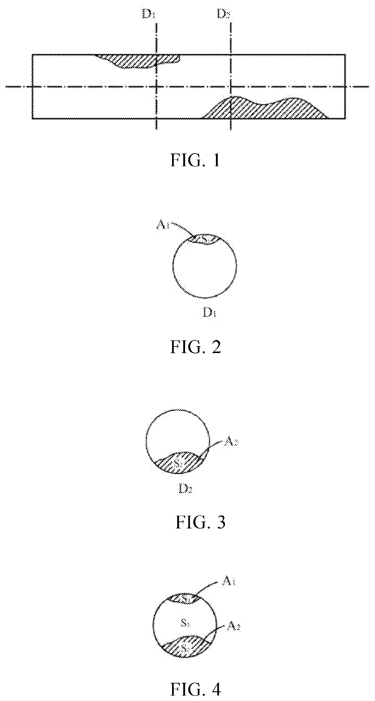
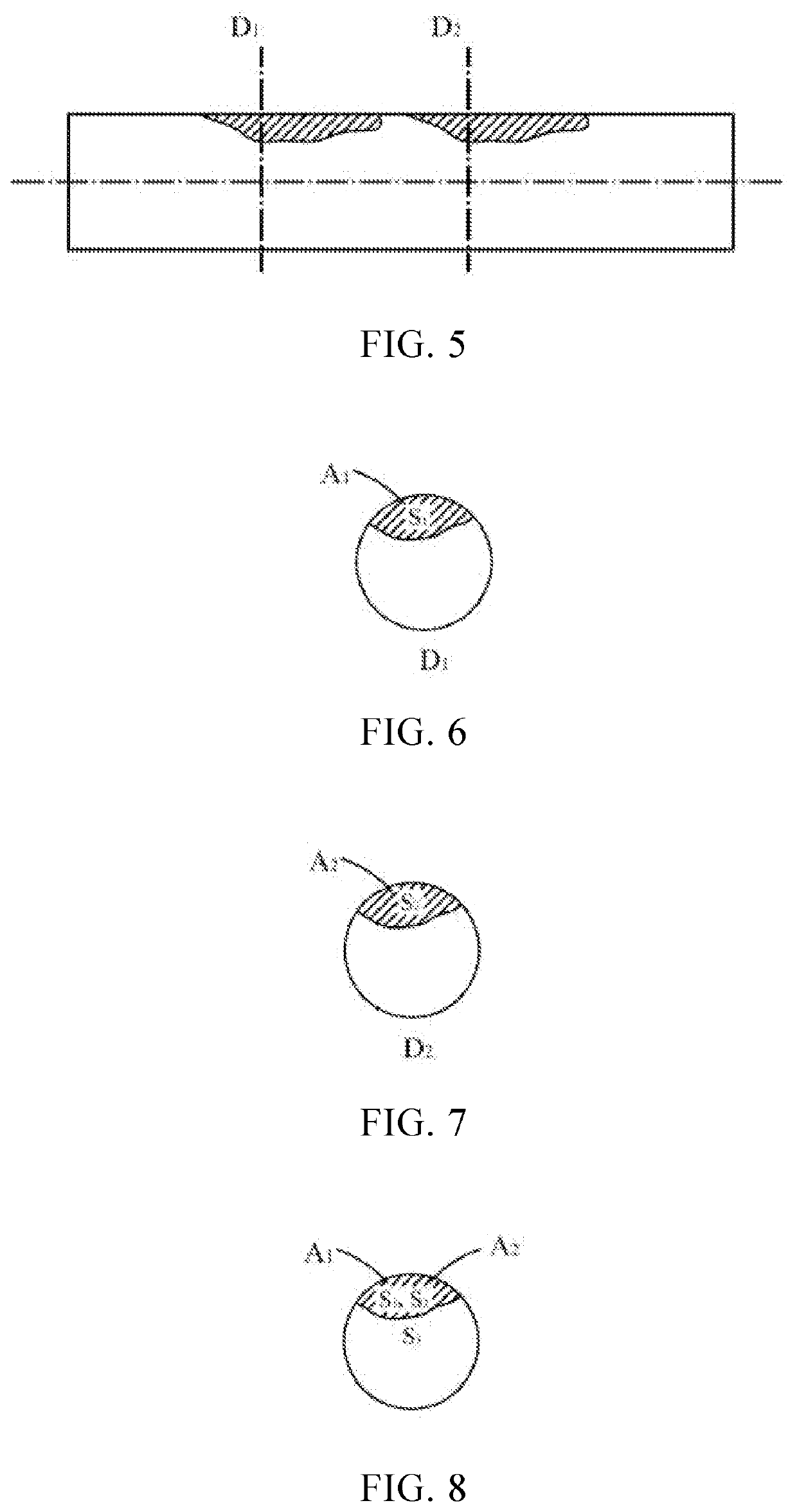
[0096]Preprocessing the geometric model to establish a cross-sectional shape model of the target blood vessel at various positions between the proximal end and the distal end;

[0097]Using the proximal end point of the target blood vessel as a reference point, the cross-sectional morphological model at different s...


PUM


Abstract

A method for obtaining vascular pressure difference includes: receiving anatomical data of a part of a blood vessel segment, obtaining a geometric model of a target blood vessel according to the anatomical data; combining individual data according to the anatomical data to obtain a blood flow model of the target blood vessel and the target blood vessel blood flow velocity V; preprocess the geometric model to establish a cross-sectional morphology model, calculate the morphological difference function f(x) of the target vessel lumen, calculate the pressure difference at any two positions of the target vessel based on morphology ΔP the difference of the target vessel lumen the relationship between function f(x) and blood flow velocity V. The method of obtaining the vascular pressure difference, by introducing the concept of morphology, the influence of the vascular morphology on the vascular pressure difference is clarified, improve the calculation accuracy of vascular pressure difference.

Description

BACKGROUND1. Technical Field[0001]The invention relates to a method and a device for obtaining vascular pressure difference, belonging to the medical technical field.2. Description of Related Art[0002]The deposition of lipids and carbohydrates in human blood on the vascular wall will form plaques on the vascular wall, which will then cause vascular stenosis; especially the vascular stenosis that occurs near the coronary artery of the heart will cause insufficient blood supply to the myocardium and induce coronary heart disease Diseases such as angina and angina pectoris pose a serious threat to human health. According to statistics, there are currently about 11 million patients with coronary heart disease in my country, and the number of patients undergoing cardiovascular interventional surgery is increasing by more than 10% every year.[0003]Although conventional medical testing methods such as coronary angiography and CT can show the severity of coronary artery stenosis, they canno...

Claims


Application Information

Patent Timeline
IPC IPC(8): A61B5/021A61B5/0285A61B5/02A61B5/00G16H50/50
CPCA61B5/021A61B5/0285G16H50/50A61B5/4887A61B5/02007A61B5/026G06T17/00G16H30/40G16H50/30G16H40/63A61B5/0066A61B6/03A61B5/1076A61B6/504A61B5/489
Inventor TU, SHENGXIANYU, WEICHEN, SHUZHANLI, YINGGUANG
Owner PULSE MEDICAL IMAGING TECH (SHANGHAI) CO LTD
Why Eureka
Try Eureka