Electrosurgical instrument

Pending Publication Date: 2021-09-30
CREO MEDICAL LTD
View PDF0 Cites 0 Cited by
  • Summary
  • Abstract
  • Description
  • Claims
  • Application Information

AI Technical Summary

Benefits of technology

The invention enables the transmission of clear images, allowing clinicians to better identify and treat areas of tissue. It also improves the accuracy of treatment. Additionally, the instrument cable has a protective sheath made of biocompatible material or coating to ensure optimal steering and stability during surgical procedures. The protective sheath has a distal portion with greater rigidity and a proximal portion with additional stiffening layers or braiding to prevent flexing or deformation.

Problems solved by technology

It is an ongoing challenge to provide both sufficient power and the additional supplies or components mentioned above in a compact enough form to fit within an instrument channel whilst maintaining flexibility and restricting power loss to acceptable (i.e. safe) levels.

Method used


Image

  • Electrosurgical instrument
  • Electrosurgical instrument
  • Electrosurgical instrument

Examples


Embodiment Construction

; FURTHER OPTIONS AND PREFERENCES

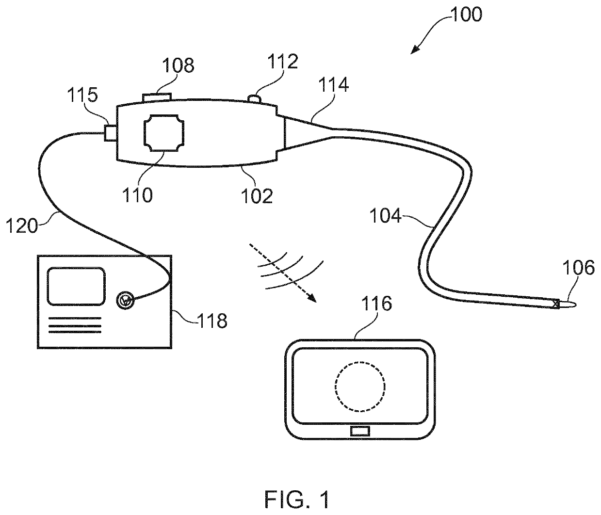
[0046]FIG. 1 is a schematic view of an electrosurgical apparatus 100 according to the present invention. The electrosurgical apparatus 100 comprises a handpiece 102 and a flexible instrument cable 104 extending away from the handpiece 102 in a distal direction. The flexible imaging cable is suitable for insertion into the body to access a treatment site. The flexible instrument cable 104 may have a biocompatible coating on its external surface so that it can be directly inserted into tissue. The instrument cable 104 may be introduced percutaneously or in a minimally invasive manner via a natural orifice. In some examples, the instrument cable 104 may be used with a separate surgical scoping device (not shown), such as a bronchoscope, endoscope, laparoscope or the like. In other examples, the imaging cable may be introduced through a guiding catheter. However, it may be particular advantageous for the imaging cable to be inserted directly (i.e. withou...


PUM


Abstract

An electrosurgical instrument for delivering radiofrequency (RF) and / or microwave energy into biological tissue that is also configured to obtain images of a treatment site performing invasive electrosurgery. In one aspect, the invention provides a chip-based images sensor at the distal end of a cable that also conveys energy for electrosurgery. In another aspect, both light emitting and light sensing element are mounted at the distal end of a probe for insertion through the instrument channel of a surgical scoping device, whereby optical signals do not need to be conveyed through the scoping device, because the transition from electrical signals to optical radiation and back to electrical signals occurs at the distal end.

Description

FIELD OF THE INVENTION[0001]The invention relates to a device for providing illumination and vision at the distal end of an electrosurgical instrument.BACKGROUND TO THE INVENTION[0002]Conventional surgical scoping devices comprise an insertion tube that can be manoeuvred to a treatment site in a patient's body via a catheter or natural orifice. The insertion tube conveys components to the treatment site. In some examples, the insertion tube comprises an observation channel for conveying an illumination signal and returning an imaging signal, and a separate instrument channel for conveying an instrument for manipulating or otherwise treating tissue at the treatment site. It can be desirable to have real-time vision of the treatment site during treatment.[0003]Electrosurgical instruments are instruments that are used to deliver radiofrequency and / or microwave frequency energy to biological tissue, for purposes such as cutting biological tissue or coagulating blood. Radiofrequency and / ...

Claims


Application Information

Patent Timeline
IPC IPC(8): A61B18/14A61B18/18A61B1/00A61B1/06A61B18/12
CPCA61B18/1492A61B18/1815A61B1/00016A61B2018/00982A61B18/1206A61B2018/1861A61B1/0684A61B18/18A61B90/361A61B90/30A61B1/313A61B1/00045A61B2090/309A61B2018/1823
Inventor HANCOCK, CHRISTOPHER PAULPRESTON, SHAUNBURN, PATRICKTAPLIN, WILLIAMJONES, AERON W.SWAIN, SANDRAULLRICH, GEORGEWEBB, DAVID
Owner CREO MEDICAL LTD
Why Eureka
Try Eureka