Process for generating genetically engineered autologous t cells

Pending Publication Date: 2021-10-21
AMGEN INC
View PDF2 Cites 0 Cited by
  • Summary
  • Abstract
  • Description
  • Claims
  • Application Information

AI Technical Summary

Benefits of technology

[0009]The invention provides a method for increasing the transgene expression in genetically engineered autologous T cells expressing a protein of interest, the method comprising inoculating a closed single use bioreactor bag containing culture media with apheresed donor cells and one or more soluble T cell activators, wherein at least one soluble T-cell activator is bound to at least one donor cell at the time of inoculation and the bioreactor bag is part of a rocking bioreactor platform, culturing the cells in the closed single use bioreactor bag continuously rocking at a rate of about 2 RPM, transducing the cells in the closed single use bioreactor bag with at least one soluble viral vector comprising a polynucleotide which encodes the protein of interest continuously rocking at a rate of about 2 RPM, and expanding the cells in the closed single use bioreactor bag at a rocking rate of about 2 RPM and increasing the culture volume and rocking the rate as needed to maintain the culture until harvest, wherein the transgene expression is greater than the transgene expression of genetically engineered autologous T cells derived from an enriched population of T cells from the same apheresed donor cells and expressing the same protein of interest. In one embodiment the apheresed donor cells comprise cells from peripheral blood. In a related embodiment the apheresed donor cells comprise nucleated and non-nucleated cells. In one embodiment the apheresed donor cells comprise leukocytes and erythrocytes. In a related embodiment the apheresed donor cells also comprise granulocytes and/or platelets. In one embodiment the apheresis is leukapheresis. In one embodiment the apheresed donor cells are washed and resuspended in a culture media. In one embodiment at least one T cell activator is an anti CD3 antibody or binding fragments thereof. In yet another embodiment the T cell activator comprises an anti CD3 antibody and an anti CD28 antibody, or binding fragments thereof. In one embodiment the T cell activator comprises at least an anti CD3 antibody, an anti CD28 antibody, and an anti CD2 antibody, or binding fragments thereof. In another embodiment the T cell activator comprises at least an anti-human CD3 monospecific tetrameric antibody complex, an anti-human CD28 monospecific tetrameric antibody complex, and an anti-human CD2 monospecific tetrameric antibody complex. In another embodiment the concentration of at least one soluble T cell activator is at least 0.001 μg/ml to at least 10 μg/ml. In a related embodiment the concentration of at least one soluble T cell activator is at least 0.1 μg/ml to at least 5 μg/ml. In another embodiment the apheresed donor cells are incubated with one or more soluble T cell activators prior to inoculating into the bioreactor bag. In a related embodiment the incubation is for a sufficient time to allow for saturation of binding of one or more soluble T cell activator to the apheresed donor cells prior to inoculation. In a related embodiment the apheresed donor cells and one or more soluble T cell activators are incubated in a transfer bag. In a related embodiment the volume of culture media in the transfer bag is about 5 ml to about 50 ml. In a related embodiment the volume of culture media in the transfer bag is about 5 ml to about 10 ml. In a related embodiment the apheresed donor cells are incubated with one or more T cell activators for at least 30 minutes or more. In a related embodiment the apheresed donor cells are incubated with one or more T cell activators for at least 1 hour. In one embodiment the number of nucleated cells within the apheresed donor cells is about 1.0E9 to about 1.3E9. In one embodiment the number of nucleated cells within the apheresed donor cells is about 1.2E9. In one embodiment the bioreactor bag is inoculated with apheresed donor cells at a cell density of about 1E6 to about 5E6 nucleated cells/ml. In a related embodiment the bioreactor bag is inoculated with apheresed donor cells at a cell density of about 2E6. In one embodiment the bioreactor bag contains at least 300 ml to at least 400 ml of culture media at inoculation. In a related embodiment the bioreactor bag contains at least 300 ml of culture media at inoculation. In one embodiment the apheresed donor cells are cultured in the bioreactor bag for about 12-24 hours. In one embodiment the culture media comprises at least one soluble cytokine. In a related embodiment the soluble cytokine selected from IL-2, IL-7, IL-15, or IL-21. In a related embodiment at least one soluble cytokine is IL-2. In a related embodiment the IL-2 is at a concentration of about 250 IU/ml to about 350 IU/ml. In a related embodiment the IL-2 is at a concentration of about 300 IU/ml. In a related embodiment the soluble cytokine is IL-7 in combination with IL-15 or IL-21. In a related embodiment the concentration of at least one cytokine is at least 5 ng/ml to at least 30 ng/ml. In a related embodiment the concentration of at least one cytokine is at least 10 ng/ml to at least 20 ng/ml. In one embodiment the culture media also comprises a WNT pathway activator. In a related embodiment the WNT pathway activator is TWS117. In a related embodiment the culture media comprises a mixture of soluble TWS117, IL-7, and IL-21. In one embodiment the culture media also comprises a soluble glycolysis inhibitor. In a related embodiment the soluble glycolysis inhibitor is 2-deoxy-D-glucose (2-DG). In one embodiment the viral vector is a retroviral vector. In one embodiment the viral vector is a lentiviral vector. In one embodiment the lentiviral vector is added at a MOI of 0.25-10. In a related embodiment the lentiviral vector is added at a MOI of 1. In one embodiment the cells are transduced for about 20-24 hours. In one embodiment following transduction, half of the culture media is removed from the bioreactor bag and replaced with an equal volume of fresh culture media. In a related embodiment the culture is incubated for about 12-24 hours. In one embodiment during expansion, fresh culture media is added to the bioreactor by fed batch/perfusion feeding and/or by perfusion. In one embodiment during expansion the culture is perfused at a rate is one bioreactor bag volume per day. In one embodiment the cells are expanded the volume of the culture media in bioreactor is incrementally increased to 1 liter during expansion. In one embodiment as the cells are expanded the volume of the culture media is incrementally increased to maintain a cell density of at least 2E6 nucleated cells/ml. In one embodiment as the cells are expanded the volume of the culture media in bioreactor is incrementally increased to 1 liter during expansion to maintain a cell density of at least 4E6 nucleated cells/ml. In one embodiment as the cells are expanded the rocking rate is incrementally increased to 6 RPM. In one embodiment at the start of expansion, the volume of culture media in the single use closed bioreactor bag is 300 ml rocking at a rate of 2 rpm at a 2° angle. In one embodiment the culture in the single use closed bioreactor bag is maintained at about 80-100% O2. In one embodiment the cells are expanded for 7 to 14 days. In one embodiment the culture, transduction, and/or expansion steps are performed at 34-37° C. In one embodiment the protein of interest is a cell surface receptor. In a related embodiment the cell surface receptor a T cell receptor, or chimeric antigen receptor. In a related embodiment the cell surface receptor recognizes an antigenic target associated with a target cell. In a related embodiment the target cell is a cancer cell. In one embodiment the genetically engineered autologous T cells are used to treat an indication in a patient in need.
[0010]The invention provides a method of treating a patient with genetically engineered autologous T

Problems solved by technology

As such, autologous cell production is more challenging than conventional biopharmaceutical processes because one lot is manufactured for one patient.
Manufacturing can only start when a patient sample is received and care must be taken that the cells are returned to the patient without significant modifications that make the cells a risk rather than an efficacious treatment.
Production is more hands on and labor intensive, and perfo

Method used


Image

  • Process for generating genetically engineered autologous t cells
  • Process for generating genetically engineered autologous t cells
  • Process for generating genetically engineered autologous t cells

Examples


Example

Example 1 Comparison Study of Three Processes

[0132]1: Autologous T cells generated with the closed continuous autologous T cell bioprocessing method where activation to expansion is conducted in a Xuri W25 Cell Expansion System. “Xuri W25 bioreactor”.

[0133]2: A bioprocessing system where activation to expansion is conducted in G-Rex® 6 and 24 well plates. “G-Rex”.

[0134]3: Hybrid bioprocessing system where activation and transduction are conducted in a gas permeable bag followed by T cell expansion in a Xuri W25 Cell Expansion System. “Permeable bag”.

[0135]In addition, two different culture media supplemented with IL2 (Media 1) or TWS119+IL7+IL21 (Media 2), were also compared using the closed continuous autologous T cell bioprocessing system and the G-Rex® plates.

[0136]T cell production was evaluated for its purity, phenotype, and in vitro properties.

[0137]On day 0, a fresh Leukopack (Hemacare, Northridge, Calif.), containing 300 ml fresh leukapheresis product collected from normal p...

Example

Example 2 
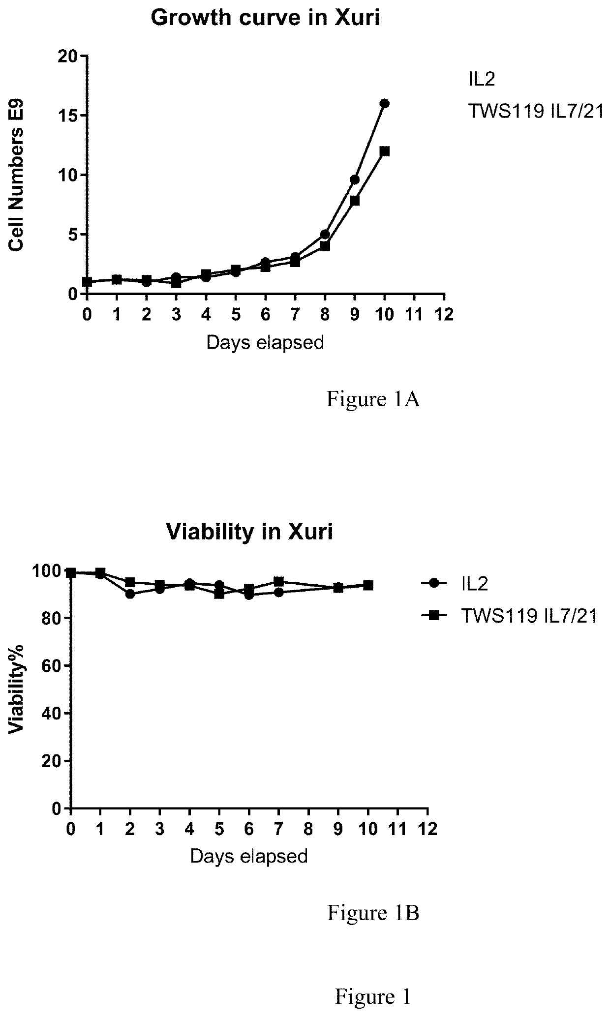
Addition of a Glycolysis Inhibitor to the Culture Media

[0165]On day 0, a fresh Leukopack (Hemacare, Northridge, Calif.), containing 300 ml fresh leukapheresis product collected from normal peripheral blood was sterile welded to a CD-600.1 Sepax Cell Separation Kit (GE Healthcare) and processed under Culture Wash-Pro program. The Leukopack® comprised leukocytes, erythrocytes, and platelets. The cells were washed in 1 L ClinMACS PBS / EDTA, 5 ml human serum albumin (Miltenyi Biotec) to remove plasma and apheresis buffers using a Sempax CPro Cell Separation System equipped with a CS-600.1 Kit (GE Healthcare), according to manufacturer's instructions. The cells were washed and counted as described above in Example 1.

[0166]A portion of the washed apheresed donor cells comprising 1.2E9 nucleated cells were transferred into two transfer bags under the Dilute program using Sepax C-Pro processing system with the same CS-600.1 Kit (a new kit was not used). Into one bag was added about...

Example

Example 3

[0185]This experiment compared CAR and TCR transduction of cells from an apheresed cell sample from a donor (Unenriched) with cells from the same apheresed cell sample that was further enriched for T cells (Enriched).

[0186]On day 0, a fresh Leukopack (Hemacare, Northridge, Calif.), containing 300 ml fresh enriched leukapheresis product collected from normal peripheral blood was sterile welded to a CD-600.1 Sepax Cell Separation Kit (GE) and processed under the Culture Wash-Pro program. The Leukopack® contained leukocytes, erythrocytes, and platelets. The cells were washed to remove plasma and apheresis buffers as described above in Example 1 and the nucleated cells were counted using NC-200™ Automated Cell Counter and split equally into two transfer bags (Sample 1, Enriched T cells) and (Sample 2, Unenriched apheresed donor cells) using the Sepax C-Pro cell processing system Dilute program.

[0187]For Sample 1, “Enriched T cells”, the T cells were isolated from the washed aph...


PUM

PropertyMeasurementUnit
Temperatureaaaaaaaaaa
Fractionaaaaaaaaaa
Timeaaaaaaaaaa

Abstract

The present invention relates to production of autologous genetically engineered T cells for use in cell therapy applications.

Description

FIELD OF DISCLOSURE[0001]The present invention relates to production of autologous genetically engineered T cells for use in cell therapy applications.BACKGROUND[0002]Adoptive T-cell therapy makes use of genetically engineered T cells and has been shown to be an effective and powerful therapeutic treatment in certain hematological indications. Autologous T cells are genetically engineered to express one or more cell surface receptors, such as chimeric antigen receptors (CARs) or T-cell receptors (TCRs), that recognize proteins of interest associated with the surface of target cells, while retaining and / or enhancing their ability to recognize and kill the target cells. In addition to the genetically engineered T cells' ability to recognize and destroy the targeted cells, successful adoptive T-cell therapy benefits from the T cells' ability to persist in the patient and continue to proliferate in response to a target antigen. Genetically engineered autologous T cells have shown great ...

Claims


Application Information

Patent Timeline
IPC IPC(8): C12N5/0783C12P21/00C12N7/00C12N15/86A61K35/17
CPCC12N5/0636C12N2740/15043C12N7/00C12N15/86A61K35/17C12N2502/1128C12N2502/1135C12N2502/115C12N2502/11C12N2501/998C12N2501/2302C12N2501/2307C12N2501/2315C12N2501/2321C12P21/00C12N2740/16043A61K39/0011C07K14/7051C12N2510/00A61K2039/5156C12P21/02
Inventor LIU, YIJUNHENCKELS, KATHRYN ANNEARBELAEZ, CARLOS
Owner AMGEN INC
Why Eureka
Try Eureka