

Method and device for measuring anatomical movement of a joint
a technology of anatomical movement and diagnostic device, which is applied in the field of diagnostic device and method for measuring the movement of a joint, can solve the problems of ignoring the concept of non-invasiveness, ignoring actually measuring the skin and surrounding tissue, so as to achieve accurate diagnostic tools and improve accuracy. the effect of movement of the join
- Summary
- Abstract
- Description
- Claims
- Application Information
AI Technical Summary
Benefits of technology
Problems solved by technology
Method used
Image

Examples
Embodiment Construction
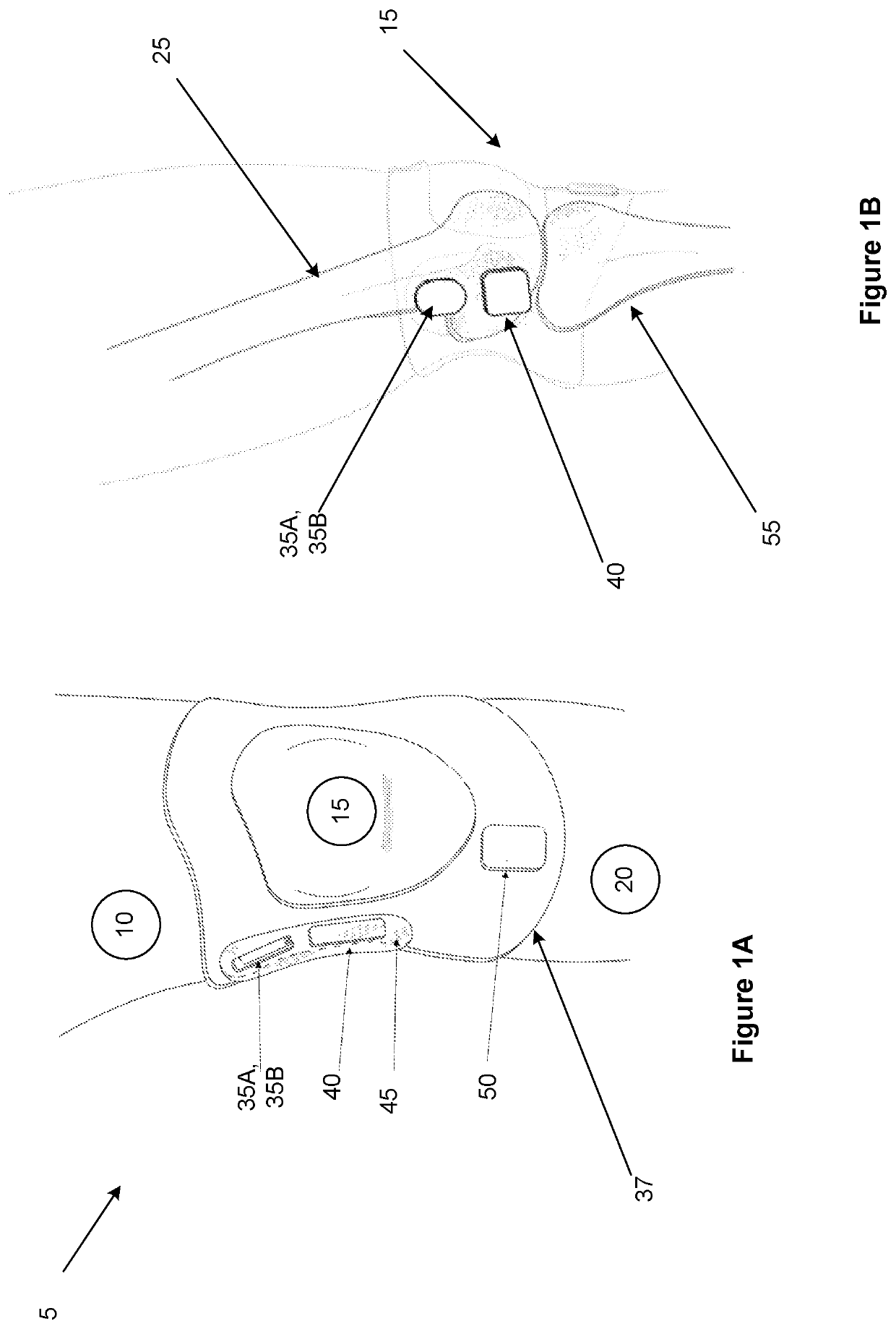
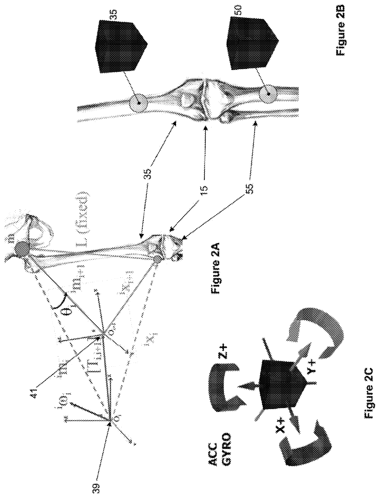
[0022]In one embodiment of the present invention, the system is directed to quantify the function of a human joint. In this embodiment, the system correlates data for 1) detecting and measuring the true anatomical bone movement in a human joint and 2) quantifying the amount of force acting through / across it. The apparatus primarily uses an inertia measurement unit (IMU) placed on the skin to detect the gross movement of the limb, with an ultrasound transducer to provide specific information on the bone movement. In a further embodiment, an electromyogram (EMG) sensor may also be used to measure the applied force applied to the joint, corresponding to the measured movement.
[0023]Advantages in using the present invention may include, in various embodiment, as follows:[0024]The ability to measure the true anatomical joint movement parameters non-invasively (including but not limited to dynamic rotational stability)[0025]Assess and compare the dynamic function between both joints (i.e. ...
PUM
 Abstract
Description
Claims
Application Information